藍藻
| シアノバクテリア門 | ||||||
|---|---|---|---|---|---|---|
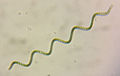
  1. (上) 糸状藍藻の1種、(下) クロオコックス属
| ||||||
| 分類 | ||||||
| ||||||
| 学名 | ||||||
| Cyanobacteria Stanier ex Cavalier-Smith, 2002 | ||||||
| 和名 | ||||||
| シアノバクテリア、藍色細菌、ラン細菌、藍藻、ラン藻、藍色植物、藍藻植物 | ||||||
| 英名 | ||||||
| cyanobacteria, cyanoprokaryotes, blue-green algae, cyanophytes | ||||||
| 下位分類[注 3] | ||||||
|
藍藻(ラン藻、らんそう、英: blue-green algae)またはシアノバクテリア[注 4] (藍色細菌、らんしょくさいきん、英: cyanobacteria)は、酸素発生を伴う光合成(酸素発生型光合成)を行う細菌の一群である。
藍藻は系統的には細菌ドメイン(真正細菌)に属する原核生物であるが、歴史的には「植物」に分類されていた(植物#リンネ以降参照)。藻類に分類されていたことから、国際細菌命名規約ではなく国際藻類・菌類・植物命名規約に基づき命名されてきた。藍藻は現在でも藻類の一員として扱われることが多いが、原核生物である点で他の藻類や陸上植物(どちらも真核生物)とは系統的に大きく異なる。しかし、陸上植物のものも含めて全ての葉緑体は細胞内共生において取り込まれた藍藻に由来すると考えられており、藍藻は植物の起源を考える上で重要な存在である。
単細胞、群体、または糸状体であり(図1)、原核生物としては極めて複雑な体をもつものもいる(→#体制)。光合成色素としてふつう青いフィコシアニンを多くもつため、クロロフィルの緑色と合わせて青緑色(藍色)をしていることが多く、学名や英名の「cyano-」はギリシア語で「青色」を意味する κυανός (kyanós) に由来する(→#光合成)。藍藻のフィコシアニンは、青い天然色素として広く利用されている(アイスキャンディーなど)。藍藻は海から淡水、陸上に広く生育し、生産者や窒素固定者として生態系において重要な役割を担っている(→#生態)。またアオコや健康食品などの形で人間生活とも密接に関わっている(→#人間との関わり)。
藍藻は、地球上に初めて現れた酸素発生型光合成生物であったと考えられている(およそ25–30億年前)。藍藻の光合成によって、地球上に初めて酸素と有機物が安定的に供給されるようになり、現在へとつながる生態系の基礎が築かれた。酸素発生型光合成というシステムは、細胞内共生(一次共生)を経て葉緑体の形で真核生物に受け継がれ、多様な真核藻類(および陸上植物)のもとともなった(→#進化)。細菌の中には、他にも光合成を行うグループが存在するが(光合成細菌と総称される)、酸素発生を伴う光合成を行うのは藍藻のみであり、他の光合成細菌は非酸素発生型[注 5]の光合成 (anoxygenic photosynthesis) を行う。
分類学的には、シアノバクテリア門(藍色細菌門、学名: Cyanobacteria)に分類される。2019年現在、メタゲノム研究(水などのサンプルから直接抽出したDNAに基づくゲノム研究であり、培養できない生物の性質を推定できる)から、藍藻に近縁であるが光合成能をもたない細菌群がいくつか見つかっている(メライナバクテリアなど)。これらの細菌群も光合成を行う藍藻とともにシアノバクテリア門に分類されることがあるが、以下では主に光合成を行う藍藻についてのみ概説する。
体制
藍藻の中には、単細胞性、群体性、糸状性の種がいる(下図2)。藍藻の多くは肉眼では判別できない微細藻であるが、群体性や糸状性の藍藻の中には、肉眼で見えるほどの大きさになるものもいる(下図6a)。

- 単細胞性 (unicellular)
- 体が1個の細胞からなる (下図3)。細胞の形は球状や桿状のものが多く(例: Synechococcus, Synechocystis)、また異極性(heteropolarity; 基端と先端で形態が異なる)を示す種もいる(例: Chamaesiphon; 下図16a)。


- 群体性 (colonial)
- 体が複数の細胞からなるが、細胞が密接していない、細胞の分化が見られないなど多細胞とは呼び難いもの(多分に伝統的な区分であり、明確な定義は難しい)(下図4)。群体全体の形態は多様である(不定形、球形、多面体、シート状、ひも状など)。また群体様式としては、多数の細胞が共通の粘液質に包まれたパルメラ状群体(palmelloid colony; 例:Aphanocapsa)が多いが、他にも細胞が密着して塊状になるサルシナ状群体(sarcinoid colony; 例:Cyanosarcina)や、分岐する粘液質の柄の先端にそれぞれ細胞が位置する樹状群体(dendroid colony)などがある。


- 糸状性 (filamentous)


細胞が密接して列状に連なっているもの (上図5)。伝統的に、糸状性の藍藻において細胞列をトリコーム(細胞糸 trichome)、1本から複数のトリコームが共通の外被に包まれたものを糸状体 (filament) とよぶ。また多数のトリコームが共通の粘液質で包まれた巨視的な群体を形成するものもいる(例: ネンジュモ属、図6)。細胞が単列 (uniseriate) に並んでいるものが多いが、多列 (multiseriate) に並んでいるものもいる(例: スチゴネマ属 Stigonema)(上図5g)。トリコームの末端の細胞はふつう他の細胞とはやや異なる形をしており (上図1g)、特に先端が肥厚している場合はカリプトラ(頂冠 calyptra)とよばれる。一部の種では、トリコームが異極性を示す(上図5d)。例えばヒゲモ属 (Rivularia) などでは、トリコームの基部に異質細胞が存在し、トリコーム先端の細胞が細長く伸びている。この場合、基部の異質細胞で窒素固定、頂端部でリン吸収を行う (つまり細胞間で形態分化とともに機能分化を示す)。トリコームは無分枝 (unbranching) であるものが多いが(上図5a, c, d)、一部は偽分枝または真分枝をする。偽分枝 (false branching) とは、トリコームの途中が分断し (ふつう細胞死による)、その一端または両端が細胞列から外れて伸長することによって形成された分枝様の構造のことである(上図5e, 図7右)。一方、真分枝 (true branching) とは、トリコームを構成する細胞が2方向以上で分裂することによって形成された分枝である(上図5b, f, g, 図7左)。分枝する種の中には、匍匐糸と直立糸の分化などの異糸性 (heterotrichous) を示すものもいる(例: Fischerella)。糸状性の藍藻は、しばしば(全てではない)細胞分化(先端の細胞、異糸性、異質細胞やアキネートなど)や細胞死を伴う形態形成(偽分枝、連鎖体形成など)、細胞間の連絡(ペリプラズムの共有やセプトソーム)を示し、原核生物ではあるものの真の多細胞体ともいえる体をもつ場合がある。
細胞
藍藻の細胞は直径1マイクロメートル (µm) 以下のこともあるが、原核生物としては大型のものが多く、直径 100 µm に達するものもいる。
細胞外被
典型的なグラム陰性細菌 (大腸菌など) と同様、藍藻の細胞壁は、ペプチドグリカン層 (peptideglycan layer) と、その外側を覆う外膜 (outer membrane) からなる。ペプチドグリカン層は、アミノ糖であるNーアセチルグルコサミンとNーアセチルムラミン酸が交互に連なった糖鎖が、オリゴペプチドで架橋された物質であるペプチドグリカン(ムレイン murein)からなる。藍藻のペプチドグリカン層は、一般的なグラム陰性細菌のそれにくらべて厚いことが多く(12–700ナノメートル (nm))、またオリゴペプチドの架橋が多い(グラム陽性細菌的な特徴)。ペプチドグリカン層が存在する細胞膜と外膜の間の空間は、ペリプラズム (periplasm) とよばれる。外膜は、細胞膜と同じく脂質二重層であるが、外側の層には糖鎖が結合した脂質(リポ多糖 lipopolysaccharide, LPS)が含まれる。他のグラム陰性細菌ではリポ多糖は毒となることがあり(内毒素)、藍藻がもつ外膜のリポ多糖にもその可能性が指摘されている。
糸状性藍藻のトリコームでは、細胞列は共通の外膜に包まれており、またペプチドグリカン層を共有している。細胞間には、ペプチドグリカン層を貫通して隣接する細胞の細胞膜同士をつなぐ連結構造であるセプトソーム(septosome、隔壁結合 septal junction、微細原形質連絡 microplasmodesmata)が多数存在する。セプトソームは長さ 25 nm、外径 15 nm、内径 6 nmほどであり、おそらく細胞間での低分子物質輸送に関与している。
外膜の外側には、結晶性の糖タンパク質からなる層が存在することがあり、S層 (surface layer, S-layer) とよばれる。このような糖タンパク質層は、細胞の保護、物質透過、接着、認識、運動、被食防御などに関与していると考えられている。


さらに細胞は、タンパク質や細胞外多糖 (exopolysaccharide) からなる細胞外高分子物質 (extracellular polymeric substance, EPS) によって覆われることがある。細胞外高分子物質は、その厚さや形態、性状に応じて、鞘(sheath; 薄く緻密で光学顕微鏡下で明瞭な構造; 上図8b, c)、夾膜 (capsule; 厚く均質で輪郭が明瞭な構造)、粘液質(slime; 細胞に沿った形を示さない不定形の構造; 上図8a)ともよばれる。多数の個体が細胞外高分子物質からなる共通の基質に包まれ、群体やバイオフィルムを形成することもある。このような外被には、無機栄養分の貯蔵、乾燥耐性、紫外線耐性、浮力増大、被食防御などの機能があると考えられている。細胞外高分子物質に含まれる紫外線吸収色素として、スキトネミン (scytonemin)やグロエオカプシン (gloeocapsin)、マイコスポリン様アミノ酸 (mycosporine-like amino acid, MAA)などが知られている。また細胞外被が石灰化(炭酸カルシウムが沈着)することがあり、おそらく光合成における二酸化炭素濃縮機構と関連している。このような石灰化によって、ストロマトライトが形成されることがある。
細胞内構造
藍藻は原核生物であり、DNAは核膜に包まれず、また葉緑体やミトコンドリア、ゴルジ体などの細胞小器官をもたない。細胞内で生体膜に包まれた構造としては、光合成における光化学反応の場であるチラコイドのみが存在する。チラコイドは扁平な袋であり、ふつう重なることなく、細胞内で同心円状(下図9)、放射状または不規則に配置する。ふつうチラコイドには、フィコビリンタンパク質からなるフィコビリソームが付着している(下図9a, 11a)。一部の藍藻(原核緑藻)はフィコビリソームを欠き、チラコイドが重なってラメラを形成している(下図9b)。藍藻では、酸素呼吸における呼吸鎖の酵素もチラコイド上に存在することがある(一部の酵素を光化学系と共有する)。最も初期に分かれた藍藻であるグロエオバクター属 (Gloeobacter) はチラコイドをもたず、光化学系は(呼吸鎖とともにパッチ状に)細胞膜上に存在する。プロクロロン属 (Prochloron) では、チラコイドの一部が膨潤して液胞状になることがある。チラコイドは、細胞膜と直接つながることはないと考えられていたが、現在では”チラコイド形成中心” (thylakoid center) が細胞膜上に存在することが示されている。光学顕微鏡下では、チラコイドが存在する細胞周縁部が色付き、チラコイドを欠く中心域が淡色に見えることがあり、伝統的に前者を有色質 (chromoplasm)、後者を中心質 (centroplasm) とよぶ。中心質にはふつうDNAが存在するため(下図9a)、この領域は核質 (nucleoplasm) ともよばれる(ただし藍藻の中には、DNAが細胞周縁部に存在する例もある)。

.jpg)

細胞内にはカルボキシソーム (carboxysome, polyhedral body) とよばれる直径200–700 nm ほどのタンパク質顆粒が存在する(上図9b, c)。カルボキシソームは主にルビスコや炭酸脱水酵素からなり、殻タンパク質で包まれている。カルボキシソームは、おそらく効率的な二酸化炭素濃縮機構に関わっており(重炭酸イオンから二酸化炭素を生成)、このため藍藻はほとんど光呼吸を示さない。ただし、おそらく特異なグリコール酸代謝経路をもつ。カルボキシソームは、炭素固定を行う他の細菌(光合成細菌や化学合成細菌)に見られることもある。
ふつう貯蔵多糖としてグリコーゲンが存在するが、α-1,6結合の分枝が少ないセミアミロペクチンやアミロースをもつものもいる。このような藍藻が貯蔵するα-グルカンは、藍藻デンプン (cyanophycean starch) ともよばれる。多くの藍藻は、アルギニンとアスパラギン酸からなる非リボソームペプチドであるシアノフィシン(下図10a)の顆粒(藍藻顆粒 cyanophycin granule)をもち、おそらく窒素貯蔵体としている(下図10b)。ただし光合成に機能するフィコビリソームを窒素貯蔵体としていることもある(窒素欠乏下ではフィコビリソームが分解され、これに由来する窒素を利用する)。細胞内には、油滴やポリリン酸体(polyphosphate body; リン貯蔵体として機能; 上図9c, 下図10c)などもふつうみられる。またβ-ヒドロキシブチレート重合体(バイオプラスチックの一種) や炭酸カルシウム(下図10c)を細胞内に貯めるものも知られている。


プランクトン性藍藻の中には、ガス胞 (gas vesicle) をもつものがいる。ガス胞は細長い小胞であり、多数のガス胞が平行に密集して"エアロトープ" (aerotope, gas vacuole) を形成している。ガス胞の膜は脂質ではなく、タンパク質からなる。この膜は水を透過しないため、ガス胞は空気で満たされ比重が軽くなり、細胞は浮くことができる。つまりガス胞は細胞中の気泡のようなものであり、水と屈折率が異なるため光学顕微鏡下では目立つ(上図10b, 下図12)。光合成産物の増加やイオン取り込みによって細胞内の膨圧が高くなるとガス胞はつぶれて細胞は沈降し、そこで光合成産物の消費やイオン排出によって膨圧が低下すると再びガス胞が膨らんで細胞は浮上する。ガス胞は藍藻に特有の構造ではなく、他のプランクトン性原核生物に見られることもある。
光合成
藍藻は、酸素発生型光合成を行う唯一の原核生物群である。藍藻は2種類の光化学系(光化学系IとII)をもつ点で、光合成を行う他の細菌(非酸素発生型光合成を行う)とは異なる。緑色硫黄細菌(クロロビウム門)やヘリオバクテリア(フィルミクテス門)は光化学系Iと相同な鉄硫黄型反応中心のみを、紅色細菌(プロテオバクテリア門)や緑色非硫黄細菌(クロロフレクサス門)は光化学系IIと相同なキノン型反応中心のみをもつ。直列した2種類の光化学系をもつことが、酸素発生型光合成(水を分解する)を可能にしていると考えられている。
知られている限り、全ての藍藻は好気環境下で酸素発生型光合成を行う。ただし、嫌気環境下で非酸素発生型光合成(光化学系Iを使用し、硫化水素を電子供与体として硫黄を生成)を行う例が知られている。また連続暗黒下でも、有機物を利用した従属栄養を行って生育可能な種(通性光独立栄養性)もいる。Synechocystis sp. PCC 6803 など従属栄養能をもつ藍藻は、光合成遺伝子の変異が致死的にならないため(光合成しなくても生きていける)、光合成研究のモデル生物として広く用いられている。またメタゲノム研究(海水などの環境から直接抽出したDNAをもとにしたゲノム解析)から、光合成能を含め代謝的に不完全(光化学系II、ルビスコ、クエン酸回路などの欠失)な藍藻 (UCYN-A, unicellular cyanobacteria group A) の存在が示されているが、これは他生物に共生して栄養的に依存して生きているものと考えられている。古くは「無色の藍藻」が報告されているが、少なくともその一部は全く別の細菌群に属することが明らかとなっている(例: ベッギアトア属)。
ほとんどの藍藻は、クロロフィル a をもつ。一部の藍藻は、クロロフィル a に加えて、クロロフィル b、d、または f をもつ。クロロフィル d や f は生物の中で一部の藍藻のみがもつ色素であり、人間の目には見えない近赤外光を光合成に利用できる。クロロフィル b(または類似色素)をもつ藍藻は、原核緑藻ともよばれる。原核緑藻のプロクロロコックス属 (Prochlorococcus) はクロロフィル a の代わりにジビニルクロロフィル a をもつ点で特異な存在であり、光合成の反応中心でジビニルクロロフィル a を用いる唯一の生物である。またアカリオクロリス属 (Acaryochloris) はクロロフィル a 量が少なく、反応中心でクロロフィル d を用いている。


ほとんどの藍藻は、光合成アンテナ色素タンパク質であるフィコビリンタンパク質をもつ。藍藻において、フィコビリンタンパク質はフィコビリソームを形成し、チラコイドに付着している。フィコビリソームの中央にはアロフィコシアニンからなるコアが位置し、そこからフィコシアニンとフィコエリスリン(後者を欠くこともある)からなるロッドが伸びている(図11a)。ふつう青色のフィコシアニンの割合が多いため、「藍藻」の名が示すように青緑色を呈する。しかし中には赤色のフィコエリスリンを多くもつため、紫色から赤色を呈する種もいる(図2, 11b)。またフィコエリスリンの代わりにフィコエリスロシアニンをもつものもいる。原核緑藻とよばれる藍藻はフィコビリンをほとんどもたないため、クロロフィルの色である緑色がそのまま見える(図11b)。
藍藻がもつカロテノイドとしては、β-カロテン、ゼアキサンチン、エキネノン、ミクソキサントフィル(ミクソール配糖体)が一般的だが、α-カロテン、カンタキサンチン、ノストキサンチン、オシラキサンチン(オシロール配糖体)などをもつものも報告されている。
藍藻の炭素固定はカルビン回路によって行われる。藍藻のもつルビスコ(リブロース1,5-ビスリン酸カルボキシラーゼ/オキシゲナーゼ)には2タイプが知られる。多くの藍藻は、緑色植物などがもつものと相同な Form IB ルビスコをもつ。このような藍藻は β-シアノバクテリア、Form IB ルビスコからなるカルボキシソームは β-カルボキシソーム とよばれる。一方、一部の藍藻(プロクロロコックス属など)は、一部のプロテオバクテリアのものと相同な Form IA ルビスコをもつ(おそらく遺伝子水平伝播による)。このような藍藻は α-シアノバクテリア、Form IA ルビスコからなるカルボキシソームは α-カルボキシソーム とよばれる。
藍藻において、酸素呼吸の電子伝達系(呼吸鎖)は細胞膜やチラコイドに存在し、後者の場合は、光合成の光化学系とタンパク質を一部共有している(プラストキノン)。また酸素呼吸におけるクエン酸回路(TCA回路)のオキソグルタル酸デヒドロゲナーゼを欠いており、この部分を別の酵素によって代謝している。
窒素固定
窒素は、タンパク質や核酸の原料として全ての生物にとって必須な元素である。窒素は窒素分子の形 (N2) で空気中に大量に存在するが、全ての真核生物を含む多くの生物は、窒素分子を直接利用することはできない。しかし原核生物の中には、窒素分子をアンモニアに変換できるものがおり、この反応は窒素固定 (nitrogen fixation) とよばれる。藍藻の中にも窒素固定が可能なものがおり、生態系において重要な役割を担っている(他の生物が利用可能な窒素栄養分の供給)。窒素を固定する酵素であるニトロゲナーゼは酸素に弱いため、酸素発生型光合成と窒素固定を1つの細胞で同時に行うことはできない。それに対応して、藍藻は以下のように光合成と窒素固定を分けて行っている。

一部の藍藻では、光が当たる日中に光合成を行い、光がない夜間に窒素固定を行う。糸状性のアイアカシオ属 (Trichodesmium) では、窒素固定を行う細胞 (diazocyte) とふつうの栄養細胞が分化しており、光合成と窒素固定を同時に異なる細胞で行うことが可能になっている。この例では細胞の形態的分化は顕著ではないが、ネンジュモ目の藍藻は、異質細胞(heterocyte, ヘテロシスト heterocyst[注 7])とよばれる形態的にも極めて特殊化した窒素固定用の細胞を形成する(上図10b, 図12)。異質細胞は光化学系の一部を欠くため細胞内に酸素が発生せず、また酸素を通さない厚い細胞壁で囲まれている。隣接する栄養細胞と接する部分では、異質細胞の細胞質は極めて細くなっており、またその部分にはときに光学顕微鏡で確認できる程の大きなシアノフィシン顆粒(極節 polar nodule)が存在する(上図10b, 図12)。異質細胞で固定された窒素はグルタミンの形で隣接細胞へ輸送され、隣接細胞からはその材料であるグルタミン酸やエネルギー源である糖(窒素固定は大量のATPを消費する)が供給される。異質細胞は通常の栄養細胞から分化するが、種によってその位置や間隔はほぼ一定であり、重要な分類形質となっている。異質細胞が分泌するペプチドによって周囲の細胞が異質細胞になることが抑制され、これによって異質細胞の間隔が一定になる例が知られている。
ゲノム

他の原核生物と同様、藍藻は環状DNAからなるゲノムをもち、また本来のゲノムDNAに加えて、小さな環状DNA(プラスミド)をもつこともある。ただし一般的な原核生物とは異なり、多くの場合ゲノム(環状DNA分子)が複数コピー存在する。多くの藍藻でゲノム塩基配列が報告されており、特に Synechocystis sp. PCC 6803 は生物として4番目、酸素発生型光合成生物として初めてゲノムが解読された。知られているものの中では、ゲノムサイズは 1.7 - 9 Mbp(Mbp = 100万塩基対)ほどであり(さらにおそらく 15 Mbp に達するものもいる)、1,700–7,000個ほどの遺伝子をもつ。この中には、一部の真核生物のゲノムより大きく多数の遺伝子をもつものもいる。
藍藻は、真核藻類にくらべて形質転換など分子生物学的解析が比較的容易なものが多く、光合成研究などのモデル生物として広く用いられている。よく用いられる藍藻として、Synechocystis sp. PCC 6803(図13)、Thermosynechococcus elongatus、Synechococcus elongatus PCC 6301、Cyanothece sp. ATCC 51142、Anabaena sp. PCC7120、Nostoc punctiforme などがある(PCC, ATCC は微生物株保存機関の略号)。
運動
藍藻は鞭毛(細菌型)をもたないが、細胞外被(S層)の糖タンパク質による遊泳運動や、線毛による匍匐運動を行うものが知られている。また藍藻において最もよく知られた運動は、多くの糸状性藍藻が示す滑走運動 (gliding movement) である。直線的な運動だけではなく、トリコームが揺れたりする運動もよく見られ(図14)、ユレモ属 (Oscillatoria) の和名、学名ともこの姿に由来する(oscillatio = 振動)。この運動は粘液多糖の噴射または糖タンパク質性外被(S層)が関係していると考えられている。
また藍藻からは、シアノバクテリオクロム (cyanobacteriochrome) やフラビン結合タンパク質、細菌型ロドプシンなどさまざまな光受容体が見つかっている。このような光受容体は、走光性や光屈性、補色馴化、細胞分化など光によって制御される現象に関わっている。
生殖


藍藻は基本的に二分裂によって増殖(図15a)、または群体や糸状体の成長を行い、群体や糸状体はその分断化によって増殖する。一部の種は、外生胞子(exospore, エクソサイト exocyte; 図16a)や内生胞子(endospore, ベオサイト baeocyte; 図16b)を形成して無性生殖を行う。
糸状性の藍藻では、ときに単純な分断化、または細胞死による隔盤 (separation disc, necridium) 形成によって糸状体の一部が切り離され、連鎖体(ホルモゴニア; hormogonium, pl. hormogonia)とよばれる短い細胞糸が形成される。連鎖体は走光性、滑走運動能、またはガス胞などをもち、散布体として機能する。
ネンジュモ目の種は、特殊化した休眠細胞であるアキネート (akinete) を形成することがある(図16c)。アキネートには異質細胞と共通する特徴(細胞壁成分、光化学系IIの非発現、スーパーオキシドジスムターゼの低活性など)があり、その分化には共通のシステムがあると考えられている。
藍藻は原核生物であり、有性生殖は行わない。ただし、外界のDNAを取り込むことや(形質転換)、ウイルス(藍藻のウイルスは特にシアノファージ cyanophage とよばれる)(図15b)によるDNA注入(形質導入)によって、遺伝子水平伝播が頻繁に起こっていることが示唆されている。
生態
藍藻は海、淡水、さらに陸上環境にも広く生育しており、藍藻がいない環境を探すのは難しい。量的にも多く、その生物量は10億トンに達するとの試算もある。一般的に、真核藻類にくらべて温度、pHが高い環境を好む傾向があるが、低温や酸性環境に生育する藍藻も少なくない。また真核藻類にくらべて、低照度で生育可能なものが多い。
貧栄養の海域(つまり沿岸域や高緯度地域を除く多くの海域)では、ピコプランクトン性(直径 2 µm 以下)の藍藻(シネココックス属 Synechococcus やプロクロロコックス属 Prochlorococcus)[注 8] が主要な生産者となっていることが多い(下図17a, b)。このような環境では、表層から真光層下部(水深 200 m 付近)まで、それぞれの光条件に適応したピコプランクトン性藍藻がすみ分けている。このようなピコプランクトン性藍藻は地球上で最も個体数が多い生物であると考えられており、シネココックス属で年平均 7.0 × 1026 細胞、プロクロロコックス属で年平均 2.9 × 1027 細胞と試算され、その純一次生産量は 12 Gt 炭素/年、海洋の全純一次生産量の25.2%に達すると推定されている。熱帯海域(特に紅海や南太平洋)では、糸状性藍藻のアイアカシオ属 (Trichodesmium) がときに多く、赤潮を形成することがある(下図17c, d)。


_contrast%C3%A9.jpg)
.JPG)
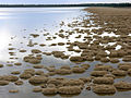
海でも淡水でも底生性の藍藻は多く、しばしばバイオフィルムを形成して基質表面を覆っている(下図19a–c)。沿岸岩礁域の潮間帯から潮上帯でも、付着性の藍藻がペンキのようにべったりと岩を覆っていることがある。またオーストラリアのシャーク湾など塩分濃度が高い浅瀬では、植食動物がいないため底生性藍藻の群落が発達し、層状構造をもつドーム状構造であるストロマトライト (stromatolite) を形成する(下図19d)。ストロマトライトに似るが、層状構造を欠くものはスロンボライト (thrombolite)、貝殻などを核に球状に形成されたものをオンコライト (oncolite) という。
.jpg)
.jpg)


藍藻は土壌や岩石表面など陸域にも多く、極地から熱帯まで広く分布している(下図20a, b)。変水性(体内の水分量が大きく変動しても、つまり乾燥しても仮死状態で生存し、再び水が得られると急速に復活する性質)による高い乾燥耐性を示すものもおり、100年近く乾燥状態に置かれたものが復活したとの報告もある。土壌表層では藍藻が土壌クラスト (soil crust) を形成し(下図20b)、土壌の安定化や窒素栄養分の供給を行うため、特に砂漠や荒野での植生遷移の初期段階において重要な働きを果たすことがある。藍藻の中には、岩石内生(岩の中に生育)のものもおり、砂漠や極地からも見つかっている。

.jpg)
.jpg)

温泉(特にアルカリ性から中性)に生育する好熱性の藍藻も知られており(温泉藻)、このような場所では温度に応じて複数種の藍藻が帯状分布を示すこともある(上図20c)。中には 70℃ 以上の高温に耐えるものや至適増殖温度が 60℃ 近いものもいる。一方、雪や氷上、北極や南極のような低温環境下に多く見られる藍藻もいる(上図20d)。氷河上で粒子状に成長した藍藻はクリオコナイト (cryoconite) とよばれ、黒っぽい色のため熱を吸収し氷河上に穴(クリオコナイトホール)を形成する。また塩湖のような塩分濃度が高い環境に生育するものもおり、浸透圧調節物質(グルコシルグリセロール、グリシン、トリメチルグリシンなど)の蓄積などによって高浸透圧に対応している。
共生
藍藻の中には、他の生物と共生しているものも少なくない。このような共生者である藍藻は、シアノビオント (cyanobiont) とよばれることがある。
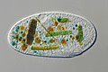
地衣類の多くは緑藻を共生者としているが、地衣の 8% 程の種は藍藻を共生者としており、特にこのような地衣は藍藻地衣 (cyanolichen) ともよばれる(下図21a)。藍藻地衣では藍藻が細胞外共生しているが、ゲオシフォン(Geosiphon; 菌根菌として重要なグループであるグロムス亜門に属する)では、藍藻 (Nostoc punctiforme) が細胞内共生している(下図21b)。これらの例では、藍藻が光合成産物(有機物)を宿主に供給し、宿主からは好適な生育環境を得ていると考えられている。同じような関係にあるものとして、海綿や等脚類、ホヤ(下図21c)、放散虫、有孔虫、繊毛虫、渦鞭毛藻(下図21d)などに藍藻が細胞外または細胞内共生している例が知られる。このような藍藻の中には、ホヤに共生するプロクロロン属 (Prochloron) のように宿主体外では生育できない絶対共生性のものもいる。


細胞内共生した藍藻が細胞小器官となった例もある。葉緑体(色素体)は、太古に細胞内共生した藍藻に起源をもつが、現在ではこの藍藻は自立能を失い、完全に宿主に制御された細胞小器官となっている。有殻糸状仮足アメーバであるビンカムリ類(Paulinella spp.; ケルコゾア門) は、葉緑体とは起源が異なる(より新しい)藍藻との細胞内共生に由来する構造(クロマトフォア chromatophore とよばれる)をもつ。この構造も既に宿主と不可分の存在であり、細胞小器官化したものであることが明らかとなっている。
_4018.JPG)
_1074.JPG)

.jpg)

水界でも、珪藻やハプト藻など光合成を行う藻類に藍藻が共生している例が知られている(上図22e)。ハフケイソウ科の珪藻に細胞内共生している藍藻は、既に自立能・光合成能を失い、楕円体 (spheroid body) とよばれる細胞小器官になっている。Braarudosphaera(ハプト藻)に共生する藍藻 (UCYN-A) も光合成能を含むいくつかの機能を欠いており、おそらく宿主に大きく依存している。
_Figure_1_lowerleft.jpg)
地衣類やサンゴにおいては、主となる共生者(それぞれ緑藻、渦鞭毛藻)とともに、窒素固定を行う藍藻が共生している例が知られている(図23)。これらの例では、光合成 (有機物供給) と窒素固定(窒素栄養分供給)を共生者の間で分業していると考えられている。
上記の例にくらべて、共生関係が明瞭ではない、より「ゆるい」共生関係も知られている。そのような例として、藍藻群集中に子嚢菌が生育しているものや、藍藻と珪藻が密集しているもの、海藻、シャジクモ類、蘚類、マングローブ植物、海草、ウキクサ、イネ、ラン(吸水根)の表面に藍藻が着生している例などが報告されている。
人間との関わり
害
富栄養湖沼において(特に夏期)、藍藻はときに大増殖してアオコ(青粉)とよばれる現象を引き起こす(下図24a; 上記参照)。アオコは様々な形で人間生活に害を与えることがある。アオコは水面に形成されるため湖沼を遮光し、水草や他の植物プランクトンの生育を妨げる。また大量に発生したアオコの夜間における呼吸、およびアオコが死んだ際の分解によって酸素が消費され、湖沼が酸欠状態になり、水生生物が死ぬことがある。一部のアオコは2-メチルイソボルネオール(下図24b)やゲオスミンなどのカビ臭物質を産生し、問題となることがある。さらにアオコを形成する藍藻の中には、下記のような藍藻毒を産生するものもいる。


藍藻の中には毒(藍藻毒、シアノトキシン cyanotoxin)を生成するものがおり、家畜やヒトに被害が生じることもある。非リボソームペプチド(リボソームにおける翻訳を介さないペプチド)であるミクロシスチン(上図24c)やノジュラリン(上図24d)はタンパク質ホスファターゼを阻害し、肝臓毒となる。またアルカロイドであるアナトキシン(上図24e)やサキシトキシンはシナプスでの伝達を阻害する神経毒となる。食用に用いられる種のスピルリナからビタミンB12の同族体が同定されたがビタミンB12の補給源にはならない、また不適切な製造工程で生産された食品での健康被害の報告が有る。
アクアリウムにおいては、水槽のガラス壁面や水草、流木などにさまざまな種の藍藻(「
食用
アフリカや中南米の湖沼で大発生する "スピルリナ"(古くは Spirulina に分類されていたためこの名でよばれるが、現在では Arthrospira に移されている)は現地では古くから食料として利用されていたが、現在では世界各地で大規模に培養され、健康食品などとして流通している(下図25a)。最大の用途は健康食品であり、錠剤などの形で市販されている (下図25b, c)。またスピルリナから抽出された光合成色素であるフィコシアニンは、水溶性の青い天然色素として冷菓などに利用されている(下図25d)。さらにカロテノイドを含むため、錦鯉の色揚剤や熱帯魚用飼料に配合されている。


他にも、食用としての藍藻の利用が世界各地で散見される。髪菜(Nostoc flagelliforme, ネンジュモ目)は中華料理の高級食材であり、内陸アジアのステップ地帯の地表に生育する(上図25e)。上記のように、このような藍藻は土壌の安定化や植生発達に重要であり、髪菜の乱獲は表土流出など環境破壊を引き起こした。そのため、中国では2000年に髪菜の採取・販売が禁止されている。髪菜に近縁の葛仙米 (Nostoc sphaeroides) やイシクラゲ (Nostoc commune) も、日本、中国、南米などで食用とされることがある。河川に生育するアシツキ (Nostoc verrucosum) は日本で古くから食用とされ、万葉集にアシツキを採取する女性を詠んだ大伴家持の歌がある。スイゼンジノリ (Aphanothece sacrum, クロオコックス目) は九州の湧水からのみ知られる藍藻であり懐石料理の高級食材などに利用され、養殖も行われている。
その他
藍藻は窒素固定能をもつため、有機肥料として用いられることがある。アカウキクサ類(薄嚢シダ類)は葉の内部に窒素固定能をもつ藍藻 (Anabaena azollae) を共生させており (上記参照)、水田の緑肥に利用されることがある。
藍藻が生成するさまざまな生理活性物質や、藍藻の細胞外被がもつ保水性、紫外線防御に関わる物質に関して、利用に向けた研究が行われている。
藍藻を利用した再生可能エネルギーの研究も盛んに行われている。例えば光合成によってエタノールを産生できる遺伝子改変藍藻、つまり光合成によって二酸化炭素を直接エタノールに変換する藍藻が作出されている。また窒素固定の際に、副産物として水素が生成されるため、これを利用した水素生産が試みられている。さらに、藍藻による光合成を直接電気に変換する研究も行われている。
米国NASAは、火星のテラフォーミングへの藍藻の利用を取り上げたことがある。
進化
ゲノムレベルでの系統解析からは、藍藻は細菌の中でクロロフレクサス門やデイノコックス・テルムス門、放線菌門、フィルミクテス門などに比較的近縁であることが示唆されており、これらを合わせてテッラバクテリア (Terrabacteria;上門レベルに相当) にまとめることが提唱されている。藍藻は、生物の進化において初めて(そして唯1回)酸素発生型光合成能を獲得した生物群であると考えられている。メタゲノム研究により、動物の腸管や土壌、汚水、地下水など様々な環境から、藍藻に近縁な非光合成細菌の系統群がいくつか見つかっている(メライナバクテリアなど)。これらの細菌が光合成能の痕跡を全くもたないことから、藍藻の祖先は嫌気性の非光合成生物であり、遺伝子の水平伝播などによって酸素発生型光合成能を獲得したと一般には考えられている。ただし、水平伝播のもとになった生物についてはよくわかっていない。
藍藻以外の光合成細菌は、藍藻がもつ光化学系Iもしくは光化学系IIのどちらか一方にのみ相同な光合成システムをもつ。これらのシステムでは水の光分解は起こらず、非酸素発生型の光合成が行われる。2つの光化学系が連結した酸素発生型の光合成を行う細菌は藍藻以外知られていない。そのため、藍藻以外の光合成細菌から、それぞれの光化学系が別個に藍藻の祖先に水平伝播したとみなされる場合が多い。しかし、各光化学系の起源と進化についてはほとんどわかっておらず、藍藻以外の光合成細菌が藍藻より以前に存在したことを示す直接的な証拠はない。そのため、藍藻のもつ2つの光化学系が別個に他の細菌に水平伝播して非酸素発生型の光合成が後から誕生した可能性も、現在のところ否定する証拠はない。実際に、緑色硫黄細菌や緑色非硫黄細菌など一部の光合成細菌については、その起源が藍藻よりもはるかに遅いことを示唆するデータが提出されており、藍藻も含めてどの光合成細菌が最も古くに誕生したのかは現在も合意に至っていない。
_(8363769094).jpg)
藍藻の進化は化石やバイオマーカー(化学化石、biomarker)、分子時計計算など様々な角度から研究が行われている。しかし、いずれも多くの異論があり、広く合意のとれた仮説は現在も存在しない。地質学的な証拠からはおよそ24億年前に地球が急速に酸化的環境に変化していったことが知られており、大酸化事変(大酸化イベント、Great Oxygenation Event, GOE)とよばれる。これは光合成によって作られた酸素が原因であるというのが最も有力な説明であり、そのため、藍藻は少なくともGOEまでには誕生していたと考えられている。しかしながら、GOE以前にも少量の酸素が存在していたことを示す証拠が複数あり、藍藻の誕生がどこまで遡るのかが議論となっている。太古代の地層に多く見られる縞状鉄鉱床は海水中の鉄などが当時すでに存在した酸素によって酸化された結果であるとする説もあるが、一方で藍藻の起源をGOEとほぼ同時期とみなす研究もある。一時期、藍藻固有のバイオマーカーが太古代から見つかり、GOE以前の藍藻および酸素の存在を示す証拠として注目されたが、その後の研究で、見つかったバイオマーカーは後世の汚染である可能性が強く、さらにバイオマーカー自体が藍藻に固有ではないことが判明している。そのほか、藍藻と考えられる化石にストロマトライトがある(図11a)。現在の地球上に見られるストロマトライトの形成にすべて藍藻が関わっていることから、ストロマトライトの化石も過去の藍藻の存在を示す証拠として扱われていたが、現在ではこれは非生物起源や、藍藻以外の光合成細菌由来のものも含まれていると考えられている。そのため、ストロマトライト単独で藍藻の進化について議論することは難しい。
いずれにせよ、藍藻の誕生によって地球環境は激変し(好気的環境、有機物安定的供給、オゾン層形成など)、現在の地球生態系の基礎が築かれた。酸素発生型光合成生物は藍藻だけである時代が長く続いたが、その後(ある研究では15億年以上前)、ある真核生物にある藍藻が細胞内共生し、やがてこの共生藍藻の増殖や代謝が宿主である真核生物に制御されるようになり、最終的に葉緑体(色素体)とよばれる細胞小器官へと変化した。この際、藍藻の細胞膜と外膜が色素体の2枚の膜になったと考えられている。この現象は一次共生 (primary endosymbiosis) とよばれ、これによって真核生物が酸素発生型光合成能を獲得した。生物の歴史の中で一次共生は唯1回の現象であったと考えられており、全ての葉緑体は単一の一次共生に由来する(その後、二次共生を経たものもある)。多数の遺伝子を用いた系統解析から、一次共生において共生者となった藍藻は、グロエオマルガリータ属 (Gloeomargarita) という淡水産単細胞性藍藻に近縁な藍藻であったことが示唆されている。
分類
古くは、藍藻は最も"原始的な植物"と考えられ、藍色植物門(藍藻植物、Cyanophyta)、藍藻綱 (Cyanophyceae)に分類されていた。しかし葉緑体の共生説が一般的となり、藍藻と他の"植物"の直接的な類縁性は認められなくなった(ただし上記のように、細胞内共生・葉緑体を通してつながっている)。現在でも藍藻は藻類の一群として扱われることが多いが、このような系統的異質性から藻類から除かれることもある(ただし藍藻を除いても藻類は多系統群である)。 これらの分類群名は植物命名規約(現 国際藻類・菌類・植物命名規約)に基づくものであり、原核生物である藍藻に対しては近年ではほとんど用いられない[注 10]。また古くは、粘藻綱 (Myxophyceae) や粘藻植物門 (Myxophta)、分裂藻綱 (Schizophyceae) という分類群名が使われていたこともある。
藍藻は原核生物であり、細菌(バクテリア、真正細菌)ドメインに属する。細菌の中では、藍藻は比較的独立した系統群を形成しており、シアノバクテリア門(藍色細菌門、学名: Cyanobacteria)として扱われる。メタゲノム研究によって見つかった、藍藻に近縁な従属栄養性細菌群である"メライナバクテリア綱" (Melainabacteria) や"セリキトクロマチア綱" (Sericytochromatia) などは、シアノバクテリア門に含めて扱われることがあるが、その場合は光合成能をもつ藍藻はオキシフォトバクテリア綱 (Oxyphotobacteria)[注 2] にまとめられ、これらの非光合成細菌群と併置される。ただし、シアノバクテリア門を光合成性のグループのみに限る考えもある。
藍藻は分類学的には細菌として扱うべきであるが、ほとんどの学名は植物命名規約(現 国際藻類・菌類・植物命名規約)に基づいて提唱されており、国際原核生物命名規約の基で提唱された学名は少ない。
クロロフィル b をもつ藍藻(原核緑藻)は、発見当初は原核緑色植物門 (Prochlorophyta) として藍藻とは別の門に分けられていた。しかしその後の研究から原核緑藻は系統的に藍藻に含まれることが示され、現在では分類群名として原核緑色植物門を用いることはない。ただし「原核緑藻 (prochlorophytes)」という語は、一般名としてはしばしば用いられる。
他の藻類と同様、藍藻はその体制(おおまかな体のつくり)に基づいて分類され、いくつかの目に分けられてきた(下表1)。細菌の分類の基準となっていた「バージェイ細菌分類便覧 (Bergey’s Manual) 第2版」でも基本的には同じ体系が用いられていた。
| 目 | バージェイ細菌分類便覧 第2版 での分類 | 特徴 | 代表属 |
|---|---|---|---|
| クロオコックス目 (Chroococcales)[注 11] | subsection I | 単細胞または群体性 | Synechococcus, Synechocystis, Chroococcus, Aphanocapsa, Coelosphaerium, Merismopedia, Microcystis |
| プレウロカプサ目 (Pleurocapsales) | subsection II | 単細胞または群体性、内生胞子 (ベオサイト) 形成 | Pleurocapsa, Chroococcidiopsis, Xenococcus |
| ユレモ目 (Oscillatoriales) | subsection III | 糸状性、異質細胞を欠く | Oscillatoria, Phormidium, Lyngbya, Arthrospira, Planktothrix, Pseudanabaena |
| ネンジュモ目 (Nostocales) | subsection IV | 糸状性 (真分枝なし)、異質細胞あり | Anabaena, Nostoc, Aphanizomenon, Cylindrospermum, Calothrix, Scytonema |
| スチゴネマ目 (Stigonematales) | subsection V | 糸状性 (真分枝あり)、異質細胞あり | Stigonema, Hapalosiphon, Fischerella |
しかし分子系統学的研究の結果、上記の分類群の多くは多系統群であることが判明しており、特に単細胞性と糸状性の間では頻繁な平行進化(特に糸状体から単細胞体への進化)が起こったと考えられている。2019年現在までに報告されている分子系統解析の結果に基づくシアノバクテリア門内の系統仮説の1つを下に示す。
| ||||||||||||||||||||||||||||||||||||||||||||||||||||||||||||||||||||||||||||||||||||||||||
| 27. 藍藻の系統仮説の一例 (基本的にゲノム塩基配列情報が明らかなもののみ): いくつかの系統解析結果に基づく. ● = 単細胞・群体 (旧クロオコックス目)、▲ = 単細胞・群体、内生胞子あり (旧プレウロカプサ目)、■ = 糸状性 (旧ユレモ目)、★ = 糸状・異質細胞あり (旧ネンジュモ目・スチゴネマ目). |
2019年現在、このような系統関係を完全に反映させた分類体系は提唱されていない。また、分子系統解析が行われた藍藻は、いまだごく一部に限られている。そのため、藍藻の分類体系構築に関しては過渡的な状況にある。2019年現在、形態形質および分子情報に基づく分類体系として、Komárek et al. (2014) をもとにした目レベルの分類体系を下表2に示す。また藍藻の分類においては、属レベルでも非単系統性(ときに下記の分類体系において複数の目に分かれるほどの)が示されているものが少なくない (Synechococcus、Synechocystis、Lyngbya など)。これらの属の一部に関しては、ゲノムレベルでの分子系統解析に基づいて多数の属に分けることが提唱されている。
表2. Komárek et al. (2014) による藍藻の分類体系 (その後に報告されたグロエオマルガリータ目を付加)
|
ギャラリー
-

-

-

-
 Gloeobacter の電子顕微鏡像
 -
 Synechococcus の透過型電子顕微鏡像
 -

-

-
 建造物表面の Gloeocapsa (黒色部)
 -
滑走運動する糸状性藍藻
-
 スピルリナ (Arthrospira platensis)
 -

-
 異極性糸状の Rivularia
 -
 異極性糸状の Gloeotrichia
 -
 スロンボライト
 -
 藍藻を捕食した Frontonia(繊毛虫)


脚注
注釈
- ^ a b c d 光合成能をもたない。シアノバクテリア門には含めないこともある。
- ^ a b この名はまだ一般的ではなく、植物命名規約に基づく「藍藻綱 (Cyanophyceae)」が暫定的に用いられることがある。
- ^ 目レベルの分類は過渡的であり、必ずしも系統を反映していない (本文参照)。
- ^ 2019年現在、高等学校の教育指導要領や、(「高等学校の生物教育における重要用語の選定について (改訂)」『学術の動向』第24巻第8号、日本学術協力財団、2019年、8_86-8_87、doi:10.5363/tits.24.8_86、ISSN 1342-3363、NAID 130007769442。)では、「シアノバクテリア」が選定されている。
- ^ 酸素非発生型とも表記される。
- ^ 補色適応 (chromatic adaptation) ともよばれる。
- ^ ヘテロシスト (heterocyst) ともよばれるが、一般的な意味でのシスト (休眠細胞) ではない。
- ^ これらの藍藻はいくつかの属に分けることが提唱されている。ただし2019年現在、これらの新しい属名は一般的ではない。
- ^ アナベナ属 (Anabaena) とされていた藍藻のうち、プランクトンとしてふつうに見られる種の多くは、ドリコスペルマム属に移されている
- ^ ただし2019年現在、原核生物の分類体系では、藍藻を分類する一般的な綱レベルの分類群名がないため、藍藻綱 (Cyanophyceae) が暫定的に用いられることがある。
- ^ 外生胞子 (エクソサイト) を形成するものはカマエシフォン目 (Chamaesiphonales) として分けられることもあった。
- ^ 一般的に「スピルリナ」とよばれる藍藻は Arthrospira に属する (この分類体系ではユレモ目)。
出典
- ^ a b c 山本純之, 磯﨑行雄「ストロマトライト研究の歴史と今後の展望」『地學雜誌』第122巻第5号、東京地学協会、2013年、791-806頁、doi:10.5026/jgeography.122.791、ISSN 0022-135X、NAID 130003385928。
- ^ a b c 巌佐庸, 倉谷滋, 斎藤成也 & 塚谷裕一 (編) (2013). “藍色細菌”. 岩波 生物学辞典 第5版. 岩波書店. pp. 1445–1446. ISBN 978-4000803144
- ^ a b c d e f 渡辺信 (1999). “藍色植物門”. In 千原光雄. バイオディバーシティ・シリーズ (3) 藻類の多様性と系統. 裳華房. pp. 160–165. ISBN 978-4785358266
- ^ "ラン藻". ブリタニカ国際大百科事典 小項目事典. コトバンクより2021年9月25日閲覧。
- ^ a b c d e f Graham, J.E., Wilcox, L.W. & Graham, L.E. (2008). “Cyanobacteria”. Algae. Benjamin Cummings. pp. 94–121. ISBN 978-0321559654
- ^ a b Büdel, B., & Kauff, F. (2012). “Prokaryotic Algae, Bluegreen Algae”. In Frey, W. (eds.). Syllabus of Plant Families. A. Engler's Syllabus der Pflanzenfamilien Part 1/1. Borntraeger. pp. 5-40. ISBN 978-3-443-01061-4
- ^ a b Garcia‐Pichel, F., Zehr, J. P., Bhattacharya, D. & Pakrasi, H. B. (2019). “What's in a name? The case of cyanobacteria”. Journal of Phycology. doi:10.1111/jpy.12934.
- ^ a b “Cyanophyceae”. Algaebase. 2021年9月23日閲覧。
- ^ a b c d e 井上勲 (2006). 藻類30億年の自然史 -藻類からみる生物進化-. 東海大学出版会. ISBN 4486017773. NCID BA75032272
- ^ “非酸素発生型光合成”. 光合成事典(Web版). 日本光合成学会 (2020年5月12日). 2022年11月11日閲覧。
- ^ 嶋田敬三「光合成細菌」『低温科学』第67巻、2009年、3-7頁。
- ^ a b c 千原光雄 (1997). “藍色植物門”. 藻類多様性の生物学. 内田老鶴圃. pp. 27–39. ISBN 978-4753640607
- ^ a b van den Hoek, C., Mann, D., Jahns, H. M. & Jahns, M. (1995). Algae: an introduction to phycology. Cambridge University Press. ISBN 978-0521316873
- ^ a b c d e f g h i Komárek, J. (2003). “Coccoid and colonial cyanobacteria”. In Wehr, J.D. & Sheath, R.G.. Freshwater Algae of North America. Ecology and Classification. Boston, MA: Academic Press. pp. 59-116. ISBN 978-0127415505
- ^ a b c d Komárek, J., Kling, H. & Komárková, J. (2003). “Filamentous cyanobacteria”. In Wehr, J.D. & Sheath, R.G.. Freshwater Algae of North America. Ecology and Classification. Boston, MA: Academic Press. pp. 117-196. ISBN 978-0127415505
- ^ Mahasneh, I.A., Grainger, S.L.J. & Whitton, B.A. (1990). “Influence of salinity on hair formation and phosphatase-activities of the blue-green-alga (cyanobacterium) Calothrix viguieri D253”. Br. Phycol. J. 25: 25-32. doi:10.1080/00071619000650021.
- ^ a b c d Flores, E. & Herrero, A. (2010). “Compartmentalized function through cell differentiation in filamentous cyanobacteria”. Nature Reviews Microbiology 8: 39-50. doi:10.1038/nrmicro2242.
- ^ Singh, S.P. & Montgomery, B.L. (2011). “Determining cell shape: adaptive regulation of cyanobacterial cellular differentiation and morphology”. Trends in Microbiology 19: 278-285. doi:10.1016/j.tim.2011.03.001.
- ^ a b c Hoiczyk, E. & Hansel, A. (2000). “Cyanobacterial cell walls: News from an unusual prokaryotic envelope”. J. Bacteriol. 182: 1191-1199. doi:10.1128/JB.182.5.1191-1199.2000.
- ^ Stewart, I., Schluter, P.J. & Shaw, G.R. (2006). “Cyanobacterial lipopolysaccharides and human health - a review”. Environ Health 5: 7. doi:10.1186/1476-069X-5-7.
- ^ Flores, E., Herrero, A., Forchhammer, K. & Maldener, I. (2016). “Septal junctions in filamentous heterocyst-forming Cyanobacteria”. Trends in Microbiology 24: 79-82. doi:10.1016/j.tim.2015.11.011.
- ^ Bornikoel, J., Carrión, A., Fan, Q., Flores, E., Forchhammer, K., Mariscal, V., ... & Maldener, I. (2017). “Role of two cell wall amidases in septal junction and nanopore formation in the multicellular cyanobacterium Anabaena sp. PCC 7120”. Frontiers in Cellular and Infection Microbiology 7: 386. doi:10.3389/fcimb.2017.00386.
- ^ Mullineaux, C. W., Mariscal, V., Nenninger, A., Khanum, H., Herrero, A., Flores, E. & Adams, D. (2008). “Mechanism of intercellular molecular exchange in heterocyst-forming cyanobacteria”. EMBO J. 27: 1299-1308. doi:10.1038/emboj.2008.66.
- ^ Šmarda, J., Šmajs, D., Komrska, J. & Krzyžánek, V. (2002). “S-layers on cell walls of cyanobacteria”. Micron 33: 257-277. doi:10.1016/S0968-4328(01)00031-2.
- ^ Ehlers, K. & Oster, G. (2012). “On the mysterious propulsion of Synechococcus”. PLoS One 7: e36081. doi:10.1371/journal.pone.0036081.
- ^ Strom, S. L., Brahamsha, B., Fredrickson, K. A., Apple, J. K. & Rodríguez, A. G. (2012). “A giant cell surface protein in Synechococcus WH8102 inhibits feeding by a dinoflagellate predator”. Environmental Microbiology 14: 807-816. doi:10.1111/j.1462-2920.2011.02640.x.
- ^ Pereira, S., Zille, A., Micheletti, E., Moradas-Ferreira, P., De Philippis, R. & Tamagnini, P. (2009). “Complexity of cyanobacterial exopolysaccharides: composition, structures, inducing factors and putative genes involved in their biosynthesis and assembly”. FEMS Microbiology Reviews 33: 917-941. doi:10.1111/j.1574-6976.2009.00183.x.
- ^ a b Plude, J.L., Parker, D.L., Schommer, O.J., Timmerman, R.J., Hagstrom, S.A., Joers, J.M. & Hnasko, R. (1991). “Chemical characterization of polysaccharide from the slime layer of the cyanobacterium Microcystis flos-aquae C3-40”. Appl. Environ. Microbiol. 57: 1696-1700.
- ^ a b De Philippis, R. & Vincenzini, M. (1998). “Exocellular polysaccharides from cyanobacteria and their possible applications”. FEMS Microbiology Reviews 22: 151-175. doi:10.1111/j.1574-6976.1998.tb00365.x.
- ^ De Philippis, R. & Vincenzini, M. (2003). “Outermost polysaccharidic investments of cyanobacteria: nature, significance and possible applications”. Recent Res. Dev. Microbiol. 7: 13-22.
- ^ a b McCarren, J. & Brahamsha, B. (2009). “Swimming motility mutants of marine Synechococcus affected in production and localization of the S-layer protein SwmA”. J. Bacteriol. 191: 1111-1114. doi:10.1128/JB.01401-08.
- ^ Reynolds, C. S. (2007). “Variability in the provision and function of mucilage in phytoplankton: facultative responses to the environment”. Hydrobiologia 578: 37-45. doi:10.1007/s10750-006-0431-6.
- ^ Sinha, R. P. & Häder D.-P. (2008). “UV-protectants in cyanobacteria”. Plant Science 174: 278-289. doi:10.1016/j.plantsci.2007.12.004.
- ^ Ehling-Schulz, M., Bilger, W. & Scherer, S. (1997). “UV-B-induced synthesis of photoprotective pigments and extracellular polysaccharides in the terrestrial cyanobacterium Nostoc commune”. J. Bacteriol. 179: 1940-1945. doi:10.1128/jb.179.6.1940-1945.1997.
- ^ Storme, J. Y., Golubic, S., Wilmotte, A., Kleinteich, J., Velázquez, D. & Javaux, E. J. (2015). “Raman characterization of the UV-protective pigment gloeocapsin and its role in the survival of cyanobacteria”. Astrobiology 15: 843-857. doi:10.1089/ast.2015.1292.
- ^ Böhm, G. A., Pfleiderer, W., Böger, P. & Scherer, S. (1995). “Structure of a novel oligosaccharide-mycosporine-amino acid ultraviolet A/B sunscreen pigment from the terrestrial cyanobacterium Nostoc commune”. J. Biol. Chem. 270: 8536-8539. doi:10.1074/jbc.270.15.8536.
- ^ Jansson, C. & Northen, T. (2010). “Calcifying cyanobacteria-the potential of biomineralization for carbon capture and storage”. Current Opinion in Biotechnology 21: 365-371. doi:10.1016/j.copbio.2010.03.017.
- ^ a b Reid, R. P., Visscher, P. T., Decho, A. W., Stolz, J. F., Bebout, B. M., Dupraz, C., Macintyre, I. G., Paerl, H. W., Pinckney, J. L., Prufert-Bebout, L., Steppe, T. F. & DesMarais, D. J. (2000). “The role of microbes in accretion, lamination and early lithification of modern marine stromatolites”. Nature 406: 989-992. doi:10.1038/35023158.
- ^ Komárek, J. & Čáslavská, J. (1991). “Thylakoidal patterns in oscillatorialean genera”. Archiv für Hydrobiologie/Algological Studies 64: 267-270.
- ^ a b Mareš, J., Strunecky, O., Bucinska, L. & Wiedermannova, J. (2019) Evolutionary patterns of thylakoid architecture in cyanobacteria. Frontiers in Microbiology 10: 277. https://doi.org/10.3389/fmicb.2019.00277
- ^ Nagarajan, A. & Pakrasi, H. B. (2001). “Membrane‐bound protein complexes for photosynthesis and respiration in cyanobacteria”. eLS: 1–8. doi:10.1002/9780470015902.a0001670.pub2.
- ^ Rippka, R. (1974). “A cyanobacterium which lacks thylakoids”. Archiv für Mikrobiologie 100: 419-436. doi:10.1007/BF00446333.
- ^ a b Cox, G. (1993). “Prochlorophyceae”. In Berner, T.. Ultrastructure of Microalgae. CRC Press. pp. 53-70. ISBN 9780849363238
- ^ Hahn, A. & Schleiff, E. (2014). “The Cell Envelope”. In Flores, E.. Cell Biology of Cyanobacteria. Caister Academic Press. pp. 29-88. ISBN 978-1-908230-92-8
- ^ Nickelsen, J. & Zerges, W. (2013). “Thylakoid biogenesis has joined the new era of bacterial cell biology”. Frontiers in Plant Science 4: 458. doi:10.3389/fpls.2013.00458.
- ^ Rast, A., Heinz, S. & Nickelsen, J. (2015). “Biogenesis of thylakoid membranes”. Biochimica et Biophysica Acta (BBA)-Bioenergetic 1847: 821-830. doi:10.1016/j.bbabio.2015.01.007.
- ^ a b Geitler, L. (1932). “Cyanophyceae”. In Rabenhorst, L.. Kryptogamen-Flora. 14. Band. Akademische Verlagsgesellschaft. pp. 1196
- ^ a b c d Price, G. D., Badger, M. R., Woodger, F. J. & Long, B. M. (2008). “Advances in understanding the cyanobacterial CO2-concentrating-mechanism (CCM): functional components, Ci transporters, diversity, genetic regulation and prospects for engineering into plants”. Journal of Experimental Botany 59: 1441-1461. doi:10.1093/jxb/erm112.
- ^ Yeates, T. O., Kerfeld, C. A., Heinhorst, S., Cannon, G. C. & Shively, J. M. (2008). “Protein-based organelles in bacteria: carboxysomes and related microcompartments”. Nature Reviews Microbiology 6: 681-691. doi:10.1038/nrmicro1913.
- ^ Colman, B. (1989). “Photosynthetic carbon assimilation and the suppression of photorespiration in the cyanobacteria”. Aquat. Bot. 34: 211-231. doi:10.1016/0304-3770(89)90057-0.
- ^ Bauwe, H., Hagemann, M. & Fernie, A. R. (2010). “Photorespiration: players, partners and origin”. Trends in Plant Science 15: 330-336. doi:10.1016/j.tplants.2010.03.006.
- ^ Codd, G.A. & Marsden, W.J.N. (1984). “The carboxysomes (polyhedral bodies) of autotrophic prokarygtes”. Biological Reviews 59: 389-422. doi:10.1111/j.1469-185X.1984.tb00710.x.
- ^ Deschamps, P., Colleoni, C., Nakamura, Y., Suzuki, E., Putaux, J. L., Buléon, A., ... & Moreira, D. (2008). “Metabolic symbiosis and the birth of the plant kingdom”. Molecular Biology and Evolution 25: 536-548. doi:10.1093/molbev/msm280.
- ^ Nakamura, Y., Takahashi, J. I., Sakurai, A., Inaba, Y., Suzuki, E., Nihei, S., ... & Kawachi, M. (2005). “Some cyanobacteria synthesize semi-amylopectin type α-polyglucans instead of glycogen”. Plant Cell Physiol. 46: 539-545. doi:10.1093/pcp/pci045.
- ^ Berg, H., Ziegler, K., Piotukh, K., Baier, K., Lockau, W. & Volkmer‐Engert, R. (2000). “Biosynthesis of the cyanobacterial reserve polymer multi‐L‐arginyl‐poly‐L‐aspartic acid (cyanophycin)”. The FEBS Journal 267: 5561-5570. doi:10.1046/j.1432-1327.2000.01622.x.
- ^ Allen, M. M. (1984). “Cyanobacterial cell inclusions”. Annual Reviews in Microbiology 38: 1-25.
- ^ a b Jensen, T. E. (1993). “Cyanobacterial ultrastructure”. In Berner, T.. Ultrastructure of Microalgae. CRC Press. pp. 7-51
- ^ Moreira, D., Tavera, R., Benzerara, K., Skouri-Panet, F., Couradeau, E., Gérard, E., Fonta, C.L., Novelo, E., Zivanovic, Y. & López-García, P. (2017). “Description of Gloeomargarita lithophora gen. nov., sp. nov., a thylakoid-bearing, basal-branching cyanobacterium with intracellular carbonates, and proposal for Gloeomargaritales ord. nov.”. International Journal of Systematic and Evolutionary Microbiology 67: 653-658. doi:10.1099/ijsem.0.001679.
- ^ Oliver, R.L. (1994). “Floating and sinking in gas-vacuolate cyanobacteria”. Journal of Phycology 30: 161-173. doi:10.1111/j.0022-3646.1994.00161.x.
- ^ Villareal, T. A. & Carpenter, E. J. (2003). “Buoyancy regulation and the potential for vertical migration in the oceanic cyanobacterium Trichodesmium”. Microb. Ecol. 45: 1-10. doi:10.1007/s00248-002-1012-5.
- ^ Walsby, A.E. (1987). “Mechanisms of buoyancy regulation by planktonic cyanobacteria with gas vesicles”. In P. Fay & C. Van Baalen. The Cyanobacteria. Elsevier. pp. 377-414
- ^ Shukla, H. D. & DasSarma, S. (2004). “Complexity of gas vesicle biogenesis in Halobacterium sp. strain NRC-1: identification of five new proteins”. Journal of Bacteriology 186: 3182-3186. doi:10.1128/JB.186.10.3182-3186.2004.
- ^ 三室守 (1999). “光合成色素にみられる多様性”. In 千原光雄. バイオディバーシティ・シリーズ (3) 藻類の多様性と系統. 裳華房. pp. 68–94. ISBN 978-4785358266
- ^ Cohen, Y., Padan, E. & Shilo, M. (1975). “Facultative anoxygenic photosynthesis in the cyanobacterium Oscillatoria limnetica”. J. Bacteriol. 123: 855-861. doi:10.1128/jb.123.3.855-861.1975.
- ^ Cohen, Y., Jorgensen, B. B., Revsbech, N. P. & Poplawski, R. (1986). “Adaptation to hydrogen sulfide of oxygenic and anoxygenic photosynthesis among cyanobacteria”. Applied and Environmental Microbiology 51: 398-407. doi:10.1128/aem.51.2.398-407.1986.
- ^ Rippka, R. (1972). “Photoheterotrophy and chemoheterotrophy among unicellular blue-green algae”. Archiv für Mikrobiologie 87: 93-98. doi:10.1007/BF00424781.
- ^ a b Zehr, J. P., Bench, S. R., Carter, B. J., Hewson, I., Niazi, F., Shi, T., ... & Affourtit, J. P. (2008). “Globally distributed uncultivated oceanic N2-fixing cyanobacteria lack oxygenic photosystem II”. Science 322: 1110-1112. doi:10.1126/science.1165340.
- ^ Tripp, H.J., Bench, S.R., Turk, K.A., Foster, R.A., Desany, B.A., Niazi, F., Affourtit, J.P. & Zehr, J.P. (2010). “Metabolic streamlining in an open-ocean nitrogen-fixing cyanobacterium”. Nature 464: 90-94. doi:10.1038/nature08786.
- ^ Kristiansen, A. (1964). “Sarcinastrum urosporae, a colourless parasitic blue-green alga”. Phycologia 4: 19-22. doi:10.2216/i0031-8884-4-1-19.1.
- ^ Lewin, R. A. & Withers, N. W. (1975). “Extraordinary pigment composition of a prokaryotic alga”. Nature 256: 735–737. doi:10.1038/256735a0.
- ^ Miyashita, H., Adachi, K., Kurano, N., Ikemoto, H., Chihara, M. & Miyachi, S. (1996). “Chlorophyll d as a major pigment”. Nature 383: 402. doi:10.1038/383402a0.
- ^ Chen, M., Schliep, M., Willows, R. D., Cai, Z. -L., Neilan, B. A. & Scheer, H. (2010). “A red-shifted chlorophyll”. Science 329: 1318-1319. doi:10.1126/science.1191127.
- ^ Chisholm, S. W., Olson, R. J., Zettler, E. R., Goericke, R., Waterbury, J. B. & Welschmeyer, N. A. (1988). “A novel free-living prochlorophyte abundant in the oceanic euphotic zone”. Nature 334: 340-343. doi:10.1038/334340a0.
- ^ Goericke, R. & Repeta, D. (1992). “The pigments of Prochlorococcus marinus: the presence of divinyl chlorophyll a and b in a marine prokaryote”. Limnology and Oceanography 37: 425-433. doi:10.4319/lo.1992.37.2.0425.
- ^ Hu, Q., Miyashita, H., Iwasaki, I., Kurano, N., Miyachi, S., Iwaki, M. & Itoh, S. (1998). “A photosystem I reaction center driven by chlorophyll d in oxygenic photosynthesis”. Proc. Natl. Acad. Sci. U.S.A. 95: 13319-13323. doi:10.1073/pnas.95.22.13319.
- ^ Sidler, W. A. (1994). “Phycobilisome and phycobiliprotein structures.”. In Sidler, W. A., & Bryant, D. A.. The Molecular Biology of Cyanobacteria. Springer, Dordrecht. pp. 139-216. ISBN 0792332229
- ^ Singh, N. K., Sonani, R. R., Rastogi, R. P. & Madamwar, D. (2015). “The phycobilisomes: an early requisite for efficient photosynthesis in cyanobacteria”. EXCLI Journal 14: 268–289. doi:10.17179/excli2014-723.
- ^ Bryant, D. A. (1982). “Phycoerythrocyanin and phycoerythrin: properties and occurrence in cyanobacteria”. Microbiology 128: 835-844. doi:10.1099/00221287-128-4-835.
- ^ a b 広瀬侑、池内昌彦、浴俊彦「シアノバクテリアの補色応答の多様性」『PLANT MORPHOLOGY』第29巻第1号、日本植物形態学会、2017年、41-45頁、doi:10.5685/plmorphol.29.41、ISSN 0918-9726、NAID 130006647120。
- ^ 巌佐庸, 倉谷滋, 斎藤成也 & 塚谷裕一 (編) (2013). “補色順化”. 岩波 生物学辞典 第5版. 岩波書店. p. 1307. ISBN 978-4000803144
- ^ Ikeuchi, M. & Ishizuka, T. (2008). “Cyanobacteriochromes: a new superfamily of tetrapyrrole-binding photoreceptors in cyanobacteria”. Photochemical & Photobiological Sciences 7: 1159-1167. doi:10.1039/B802660M.
- ^ 光合成事典. 日本光合成学会編.
- ^ Takaichi, S. & Mochimaru, M. (2007). “Carotenoids and carotenogenesis in cyanobacteria: Unique ketocarotenoids and carotenoid glycosides”. Cell Mol. Life Sci. 64: 2607-2619. doi:10.1007/s00018-007-7190-z.
- ^ Scherer, S., Almon, H. & Böger, P. (1988). “Interaction of photosynthesis, respiration and nitrogen fixation in cyanobacteria”. Photosynthesis Research 15: 95-114. doi:10.1007/BF00035255.
- ^ Zhang, S. & Bryant, D. A. (2011). “The tricarboxylic acid cycle in cyanobacteria”. Science 334: 1551-1553. doi:10.1126/science.1210858.
- ^ Stewart, W.D.P. (1980). “Some aspects of structure and function in N fixing cyanobacteria”. Annual Reviews in Microbiology 34: 497-536. doi:10.1146/annurev.mi.34.100180.002433.
- ^ Berman-Frank, I., Lundgren, P. & Falkowski, P. (2003). “Nitrogen fixation and photosynthetic oxygen evolution in cyanobacteria”. Res. Microbiol. 154: 157-164. doi:10.1016/S0923-2508(03)00029-9.
- ^ Díez, B., Bergman, B. & El-Shehawy, R. (2008). “Marine diazotrophic cyanobacteria: out of the blue”. Plant Biotechnol. 25: 221-225. doi:10.5511/plantbiotechnology.25.221.
- ^ Rippka, R., Deruelles, J., Waterbury, J. B., Herdman, M. & Stanier, R. Y. (1979). “Generic assignments, strain histories and properties of pure cultures of cyanobacteria”. Microbiology 111: 1-61. doi:10.1099/00221287-111-1-1.
- ^ León, C., Kumazawa, S. & Mitsui, A. (1986). “Cyclic appearance of aerobic nitrogenase activity during synchronous growth of unicellular cyanobacteria”. Current Microbiology 13: 149-153. doi:10.1007/BF01568510.
- ^ El-Shehawy, R., Lugomela, C., Ernst, A. & Bergman, B. (2003). “Diurnal expression of hetR and diazocyte development in the filamentous non-heterocystous cyanobacterium Trichodesmium erythraeum”. Microbiology 149: 1139-1146. doi:10.1099/mic.0.26170-0.
- ^ a b Bergman, B., Sandh, G., Lin, S., Larsson, J. & Carpenter, E. J. (2013). “Trichodesmium - a widespread marine cyanobacterium with unusual nitrogen fixation properties”. FEMS Microbiology Reviews 37: 286-302. doi:10.1111/j.1574-6976.2012.00352.x.
- ^ “藍藻の分類”. 浮遊性藍藻データベース. 国立科学博物館. 2021年9月25日閲覧。
- ^ Wolk, C.P., Ernst, A., Elhai, J. (1994). “Heterocyst metabolism and development”. In Bryant, D.A.. The Molecular Biology of Cyanobacteria. Kluwer Academic Publishers. pp. 769-823. ISBN 0792332229
- ^ Kumar, K., Mella-Herrera, R. A. & Golden, J. W. (2010). “Cyanobacterial heterocysts”. Cold Spring Harbor Perspectives in Biology 2: a000315. doi:10.1101/cshperspect.a000315.
- ^ Marco, G., Lange, C. & Soppa, J. (2011). “Ploidy in cyanobacteria”. FEMS Microbiology Letters 323: 124-131. doi:10.1111/j.1574-6968.2011.02368.x.
- ^ Kaneko, T., Sato, S., Kotani, H., Tanaka, A., Asamizu, E., Nakamura, Y., ... & Kimura, T. (1996). “Sequence analysis of the genome of the unicellular cyanobacterium Synechocystis sp. strain PCC6803. II. Sequence determination of the entire genome and assignment of potential protein-coding regions”. DNA Research 3: 109-136. doi:10.1093/dnares/3.3.109.
- ^ Herdman, M., Janvier, M., Rippka, R. & Stanier, R. Y. (1979). “Genome size of Cyanobacteria”. Journal of General Microbiology 111: 73-85. doi:10.1099/00221287-111-1-73.
- ^ 広瀬侑、佐藤桃子、池内昌彦「シアノバクテリア (光合成研究法) -- (植物・藻類・細菌の材料の入手と栽培・培養)」『低温科学』第67巻、北海道大学低温科学研究所、2008年、9-15頁、ISSN 18807593、NAID 120001492974。
- ^ a b Duggan, P. S., Gottardello, P. & Adams, D. G. (2007). “Molecular analysis of genes involved in pilus biogenesis and plant infection in Nostoc punctiforme”. J. Bacteriol. 189: 4547-4551. doi:10.1128/JB.01927-06.
- ^ Jarrell, K.F. & McBride, M.J. (2008). “The surprisingly diverse ways that prokaryotes move”. Nature Reviews Microbiology 6: 466-476. doi:10.1038/nrmicro1900.
- ^ Montgomery, B. L. (2007). “Sensing the light: photoreceptive systems and signal transduction in cyanobacteria”. Molecular Microbiology 64: 16-27. doi:10.1111/j.1365-2958.2007.05622.x.
- ^ 広瀬侑、池内昌彦「シアノバクテリアの補色順化における光色感知機構」『化学と生物』第54巻第6号、日本農芸化学会、2016年、403-407頁、doi:10.1271/kagakutoseibutsu.54.403、ISSN 0453-073X、NAID 130006772575。
- ^ Anagnostidis, K. & Komárek, J. (1988). “Modern approach to the classification system of cyanophytes. 3. Oscillatoriales”. Archiv für Hydrobiologie/Algological Studies 50/53: 327-472.
- ^ Meeks, J. C. & Elhai, J. (2002). “Regulation of cellular differentiation in filamentous cyanobacteria in free-living and plant-associated symbiotic growth states”. Microbiology and Molecular Biology Reviews 66: 94-121. doi:10.1128/MMBR.66.1.94-121.2002.
- ^ Kaplan-Levy, R. N., Hadas, O., Summers, M. L., Rücker, J., & Sukenik, A. (2010). “Akinetes: dormant cells of cyanobacteria”. Dormancy and Resistance in Harsh Environments. Springer Berlin Heidelberg. pp. 5-27. ISBN 978-3-642-12421-1.
- ^ Zhang, C.-C., Laurent, S., Sakr, S., Peng, L. & Bédu, S. (2006). “Heterocyst differentiation and pattern formation in cyanobacteria: a chorus of signals.”. Mol. Microbiol. 59: 367-375. doi:10.1111/j.1365-2958.2005.04979.x.
- ^ a b Coleman, M. L., Sullivan, M. B., Martiny, A. C., Steglich, C., Barry, K., DeLong, E. F. & Chisholm, S. W. (2006). “Genomic islands and the ecology and evolution of Prochlorococcus”. Science 311: 1768-1770. doi:10.1126/science.1122050.
- ^ a b c Whitton, B.A. & Potts, M. (2000). The Ecology of Cyanobacteria: Their Diversity in Time and Space. Kluwer Academic Pub.. pp. 669. ISBN 0-09-941464-3
- ^ a b c Whitton, B.A., ed (2012). Ecology of Cyanobacteria II: Their Diversity in Space and Time. Springer Science & Business Media. ISBN 978-94-007-3854-6
- ^ Garcia-Pichel, F., Belnap, J., Neuer, S. & Schanz, F. (2003). “Estimates of global cyanobacterial biomass and its distribution”. Algological Studies 109: 213-227. doi:10.1127/1864-1318/2003/0109-0213.
- ^ a b c Castenholz, R.W. & Waterbury, J.B. (1989). “Oxygenic photosynthetic bacteria. Group I. Cyanobacteria”. Bergey’s Manual of Systematic Bacteriology 3: 1710-1789.
- ^ a b Quesada, A. & Vincent, W. F. (2012). “Cyanobacteria in the cryosphere: snow, ice and extreme cold”. Ecology of Cyanobacteria II. Springer Net.. pp. 387-399. ISBN 978-94-007-3854-6.
- ^ Steinberg, C.E.W., Schäfer, H., Beisker, W., Brüggemann, R. (1998). “Deriving restoration goals for acidified lakes from taxonomic studies”. Restor, Ecol. 6: 327-335. doi:10.1046/j.1526-100X.1998.06403.x.
- ^ van Liere, L. & Walsby, A.E. (1982). “Interactions of cyanobacteria with light”. In Carr, N.G. and Whitton, B.A.. The Biology of the Cyanobacteria. Blackwell Science Publications. pp. 9-45. ISBN 0-520-04717-6.
- ^ a b c Walter, J. M., Coutinho, F. H., Dutilh, B. E., Swings, J., Thompson, F. L. & Thompson, C. C. (2017). “Ecogenomics and taxonomy of Cyanobacteria phylum”. Frontiers in Microbiology 8: 2132. doi:10.3389/fmicb.2017.02132.
- ^ Weisse, T. (1993). “Dynamics of autotrophic picoplankton in marine and freshwater ecosystems”. In Jones, J.G.. Advances in Microbial Ecology, Vol. 13. Plenum Press. pp. 327-370. doi:10.1007/978-1-4615-2858-6_8.
- ^ Veldhuis, M.J.W., Kraay, G.W., van Bleijswijk, J.D.L. & Baars, M.A. (1997). “Seasonal and spatial variability in phytoplankton biomass, productivity and growth in the northwestern Indian ocean: the southwest and northeast monsoon, 1992-1993”. Deep Sea Research Part I: Oceanographic Research Papers 44: 425-449. doi:10.1016/S0967-0637(96)00116-1.
- ^ Zwirglmaier, K., Jardillier, L., Ostrowski, M., Mazard, S., Garczarek, L., Vaulot, D., Not, F., Massana, R., Utioa, O. & Scanlan, D. J. (2008). “Global phylogeography of marine Synechococcus and Prochlorococcus reveals a distinct partitioning of lineages among oceanic blooms”. Environ. Microbiol. 10: 147-161. doi:10.3389/fmicb.2018.01393.
- ^ Flombaum, P., Gallegos, J. L., Gordillo, R. A., Rincón, J., Zabala, L. L., Jiao, N., ... & Vera, C. S. (2013). “Present and future global distributions of the marine Cyanobacteria Prochlorococcus and Synechococcus”. Proc. Natl. Acad. Sci. U.S.A. 110: 9824-9829. doi:10.1073/pnas.1307701110.
- ^ Callieri, C. (2007). “Picophytoplankton in freshwater ecosystems: the importance of small-sized phototrophs”. Freshwater Reviews 1: 1-28. doi:10.1608/FRJ-1.1.1.
- ^ “ネンジュモ目”. 浮遊性藍藻データベース. 国立科学博物館. 2021年9月25日閲覧。
- ^ a b 渡辺真利代; 原田健一; 藤木博太 (編) (1994). アオコ : その出現と毒素. 東京大学出版会. pp. 257. ISBN 4-13-066152-3. NCID BN11097702
- ^ Manage, P.M., Kawabata, Z. & Nakano, S. (2001). “Dynamics of cyanophage-like particles and algicidal bacteria causing Microcystis aeruginosa mortality”. Limnology 2: 73-78. doi:10.1007/s102010170002.
- ^ Sukenik, A., Eshkol, R., Livne, A., Hadas, O., Rom, M., Tchernov, D., Vardi, A. & Kaplan, A. (2002). “Inhibition of growth and photosynthesis of the dinoflagellate Peridinium gatunense by Microcystis sp. (cyanobacteria): a novel allelopathic mechanism”. Limnol. Oceanogr. 47: 1656-1663. doi:10.4319/lo.2002.47.6.1656.
- ^ Mizuta, S., Imai, H., Chang, K.-H., Doi, H., Nishibe, Y. & Nakano, S. (2010). “Grazing on Microcystis (Cyanophyceae) by testate amoebae with special reference to cyanobacterial abundance and physiological state”. Limnplogy 12: 205-211. doi:10.1007/s10201-010-0341-1.
- ^ Zotina, T., Köster, O. & Jüttner, F. (2003). “Photoheterotrophy and light‐dependent uptake of organic and organic nitrogenous compounds by Planktothrix rubescens under low irradiance”. Freshwater Biology 48: 1859-1872. doi:10.1046/j.1365-2427.2003.01134.x.
- ^ a b Tang, E. P. Y., Tremblay, R. & Vincent, W. F. (1997). “Cyanobacterial dominance of polar freshwater ecosystems: are high-latitude mat-formers adapted to low temperature?”. J. Phycol. 33: 171-181. doi:10.1111/j.0022-3646.1997.00171.x.
- ^ Shapiro, R. S. (2000). “A comment on the systematic confusion of thrombolites”. Palaios 15 (2): 166-169. doi:10.2307/3515503.
- ^ Corsetti, F. A., Awramik, S. M. & Pierce, D. (2003). “A complex microbiota from snowball Earth times: microfossils from the Neoproterozoic Kingston Peak Formation, Death Valley, USA”. Proc. Natl. Acad. Sci. U.S.A. 100: 4399-4404. doi:10.1073/pnas.0730560100.
- ^ Ward, D. M. & Castenholz, R. W. (2000). “Cyanobacteria in geothermal habitats”. The Ecology of Cyanobacteria. Springer Netherlands. pp. 37-59. ISBN 0-09-941464-3
- ^ Wierzchos, J., Ascaso, C. & McKay, C. P. (2006). “Endolithic cyanobacteria in halite rocks from the hyperarid core of the Atacama Desert”. Astrobiology 6: 415-422. doi:10.1089/ast.2006.6.415.
- ^ 竹内望「クリオコナイトと氷河の暗色化」『低温科学』第70巻、北海道大学低温科学研究所、2012年、165-172頁、ISSN 1880-7593、NAID 40019324597。
- ^ Fulda, S., Mikkat, S., Schroder, W., Hagemann, M. (1999). “Isolation of salt-induced periplasmic proteins from Synechocystis sp. strain PCC 6803”. Arch. Microbiol. 171: 214-217. doi:10.1007/s002030050702.
- ^ a b c Adams, D. G. (2000). “Symbiotic interactions”. In Whitton, B.A. & Potts, M.. Ecology of Cyanobacteria: Their Diversity in Time and Space. Kluwer Academic Publishers. pp. 523-561. ISBN 0-09-941464-3
- ^ a b c d Adams, D. G., Duggan, P. S. & Jackson, O. (2012). “Cyanobacterial symbioses”. In Whitton, B.A.. Ecology of Cyanobacteria II: Their Diversity in Space and Time. Springer Science+Business Media B.V.. pp. 593-675. ISBN 978-94-007-3854-6
- ^ a b c Adams, D. G., Bergman, B., Nierzwicki-Bauer, S. A., Rai, A. N. & Schüßler, A. (2006). “Cyanobacterial-plant symbioses”. In Dworkin M, Falkow S, Rosenberg E, Schleifer K-H, Stackebrandt E. The Prokaryotes. A Handbook on the Biology of Bacteria, vol 1, 3rd ed. Symbiotic Associations, Biotechnology, Applied Microbiology. Springer. pp. 331-363. ISBN 978-1-4757-2193-5
- ^ a b Carpenter, E.J. (2002). “Marine cyanobacterial symbioses”. Biol. Environ. Proc. R Ir Acad. 102B: 15-18. doi:10.1007/0-306-48005-0_2.
- ^ Paerl, H. (1992). “Epi- and endobiotic interactions of cyanobacteria”. In Reisser, W.. Algae and Symbioses: Plants, Animals, Fungi, Viruses, Interactions Explored. Biopress Limited. pp. 537-565
- ^ Decelle, J., Colin, S. & Foster, R. A. (2015). “Photosymbiosis in marine planktonic protists”. Marine Protists. Springer Japan. pp. 465-500. doi:10.1007/978-4-431-55130-0_19
- ^ a b Rikkinen, J. (2002). “Cyanolichens: an evolutionary overview”. In Rai, A.N., Bergman, B. & Rasmussen, U.. Cyanobacteria in Symbiosis. Kluwer Academic Publishers, Dordrecht. pp. 31-72. ISBN 1-4020-0777-9
- ^ Gehrig, H., Schüßler, A. & Kluge, M. (1996). “Geosiphon pyriforme, a fungus forming endocytobiosis withNostoc (Cyanobacteria), is an ancestral member of the glomales: evidence by SSU rRNA analysis”. Journal of Molecular Evolution 43: 71-81. doi:10.1007/BF02352301.
- ^ Mollenhauer, D., Mollenhauer, R. & Kluge, M. (1996). “Studies on initiation and development of the partner association in Geosiphon pyriforme (Kütz.) v. Wettstein, a unique endocytobiotic system of a fungus (Glomales) and the cyanobacterium Nostoc punctiforme (Kütz.) Hariot”. Protoplasma 193: 3-9. doi:10.1007/BF01276630.
- ^ Schüßler, A. & Wolf, E. (2005). “Geosiphon pyriformis - a Glomeromycotan soil fungus forming endosymbiosis with Cyanobacteria”. In Vitro Culture of Mycorrhizas. Soil Biology, Volume 4, Part V. pp. 271-289. ISBN 3-540-24027-6
- ^ Usher, K.M. (2008). “The ecology and phylogeny of cyanobacterial symbionts in sponges”. Marine Ecology 29: 178-192. doi:10.1111/j.1439-0485.2008.00245.x.
- ^ Lindquist, N., Barber, P.H. & Weisz, J.B. (2005). “Episymbiotic microbes as food and defence for marine isopods: unique symbioses in a hostile environment”. Proc. R Soc. Lond. B 272: 1209-1216. doi:10.1098/rspb.2005.3082.
- ^ Münchhoff, J., Hirose, E., Maruyama, T., Sunairi, M., Burns, B.P., & Neilan, B.A. (2007). “Host specificity and phylogeography of the prochlorophyte Prochloron sp., an obligate symbiont in didemnid ascidians”. Environ. Microbiol. 9: 890-899. doi:10.1111/j.1462-2920.2006.01209.x.
- ^ a b c Foster, R. A. Carpenter, E. J. & Bergman, B. (2006). “Unicellular cyanobionts in open ocean dinoflagellates, radiolarians, and tintinnids: ultrastructural characterization and immuno-localization of phycoerythrin and nitrogenase”. Journal of Phycology 42: 453-463. doi:10.1111/j.1529-8817.2006.00206.x.
- ^ a b c Foster, R. A., Collier, J. L. & Carpenter , E. J. (2006). “Reverse transcription PCR amplification of cyanobacterial symbiont 16S rRNA sequences from single non-photosynthetic eukaryotic marine planktonic host cells”. Journal of Phycology 42: 243-250. doi:10.1111/j.1529-8817.2006.00185.x.
- ^ Lee, J.J. (2006). “Algal symbiosis in larger foraminifera”. Symbiosis 42: 63-75.
- ^ Escalera, L., Reguera, B., Takishita, K., Yoshimatsu, S., Koike, K. & Koike, K. (2011). “Cyanobacterial endosymbionts in the benthic dinoflagellate Sinophysis canaliculata (Dinophysiales, Dinophyceae)”. Protist 162: 304-314. doi:10.1016/j.protis.2010.07.003.
- ^ Jyothibabu, R., Madhu, N.V., Maheswaran, P.A., Devi, C.R.A., Balasubramanian, T., Nair, K.K.C. & Achuthankutty, C.T. (2006). “Environmentally-related seasonal variation in symbiotic associations of heterotrophic dinoflagellates with cyanobacteria in the western Bay of Bengal”. Symbiosis 42: 51-58.
- ^ a b Archibald, J.M. (2009). “The puzzle of plastid evolution”. Curr. Biol. 19: R81-88. doi:10.1016/j.cub.2008.11.067.
- ^ 中山卓郎、石田健一郎「もう一つの一次共生?-Paulinella chromatophora とそのシアネレ―」『原生動物学雑誌』第41巻第1号、日本原生生物学会、2008年、27-31頁、doi:10.18980/jjprotozool.41.1_27、ISSN 0388-3752、NAID 130006070219。
- ^ Rai, A. N., Söderbäck, E. & Bergman, B. (2000). “Cyanobacterium-plant symbioses”. New Phytologist 147: 449-481. doi:10.1046/j.1469-8137.2000.00720.x.
- ^ a b Adams, D. G. & Duggan, P. S. (2008). “Cyanobacteria-bryophyte symbioses”. J. Exp. Bot. 59: 1047-1058. doi:10.1093/jxb/ern005.
- ^ Peters, G.A. (1991). “Azolla and other plant-cyanobacteria symbioses - aspects of form and function”. Plant Soil 137: 25-36. doi:10.1007/BF02187428.
- ^ a b Papaefthimiou, D., Van Hove, C., Lejeune, A., Rasmussen, U. & Wilmotte, A. (2008). “Diversity and host specificity of genus Azolla cyanobionts”. J. Phycol. 44: 60-70. doi:10.1111/j.1529-8817.2007.00448.x.
- ^ Costa, J.-L. & Lindblad, P. (2003). “Cyanobacteria in symbiosis with cycads”. In Rai, A.N., Bergman, B. & Rasmussen, U.. Cyanobacteria in Symbiosis. Kluwer Academic Publishers, Dordrecht. pp. 195-205. ISBN 1-4020-0777-9
- ^ Bergman, B. (2002). “The Nostoc-Gunnera symbiosis”. In Rai, A.N., Bergman, B. & Rasmussen, U.. Cyanobacteria in Symbiosis. Kluwer Academic Publishers. pp. 207-232. ISBN 1-4020-0777-9
- ^ Bergman, B. & Osborne, B. (2002). “The Gunnera-Nostoc symbiosis”. Biol. Environ. Proc. R Ir Acad. 102B: 35-39.
- ^ Cox, P.A., Banack, S.A. & Murch, S.J. (2003). “Biomagnification of cyanobacterial neurotoxins and neurodegenerative disease among the Chamorro people of Guam”. Proc. Natl. Acad. Sci. U.S.A. 100: 13380-13383. doi:10.1073/pnas.2235808100.
- ^ Jahson, S., Rai, A. N. & Bergman, B. (1995). “Intracellular cyanobiont Richelia intracellularis: ultrastructure and immuno-localisation of phycoerythrin, nitrogenase, Rubisco and glutamine synthetase”. Marine Biology 124: 1-8. doi:10.1007/BF00349140.
- ^ Foster, R. A. & Zehr, J. P. (2006). “Characterization of diatom-cyanobacteria symbioses on the basis of nifH, hetR and 16S rRNA sequences”. Environ. Microbiol. 8: 1913-1925. doi:10.1111/j.1462-2920.2006.01068.x.
- ^ Foster, R.A., Kuypers, M.M.M., Vagner, T., Paerl, R.W., Muzat, N. & Zehr, J.P. (2011). “Nitrogen fixation and transfer in open ocean diatom-cyanobacterial symbioses”. ISME J. 5: 1484-1493. doi:10.1038/ismej.2011.26.
- ^ Foster, R.A., Subramaniam, A. & Zehr, J.P. (2009). “Distribution and activity of diazotrophs in the Eastern Equatorial Atlantic”. Environ. Microbiol. 11: 741-750. doi:10.1111/j.1462-2920.2008.01796.x.
- ^ White, A.E., Prahl, F.G., Letelier, R.M. & Popp, B.N. (2007). “Summer surface waters in the Gulf of California: prime habitat for biological nitrogen fixation”. Glob. Biogeochem. Cycles 21: GB2017. doi:10.1029/2006GB002779.
- ^ Hagino, K., Onuma, R., Kawachi, M. & Horiguchi, T. (2013). “Discovery of an endosymbiotic nitrogen-fixing cyanobacterium UCYN-A in Braarudosphaera bigelowii (Prymnesiophyceae)”. PLoS One 8: e81749. doi:10.1371/journal.pone.0081749.
- ^ Thompson, A., Carter, B. J., Turk‐Kubo, K., Malfatti, F., Azam, F. & Zehr, J. P. (2014). “Genetic diversity of the unicellular nitrogen‐fixing cyanobacteria UCYN‐A and its prymnesiophyte host”. Environmental Microbiology 16: 3238-3249. doi:10.1111/1462-2920.12490.
- ^ Kneip, C., Voß, C., Lockhart, P. J. & Maier, U. G. (2008). “The cyanobacterial endosymbiont of the unicellular algae Rhopalodia gibba shows reductive genome evolution”. BMC Evol. Biol. 8: 30. doi:10.1111/1462-2920.12490.
- ^ Lesser, M. P., Mazel, C. H., Gorbunov, M. Y. & Falkowski, P. G. (2004). “Discovery of symbiotic nitrogen-fixing cyanobacteria in corals”. Science 305: 997-1000. doi:10.1126/science.1099128.
- ^ Lesser, M.P., Falcón, L.I., Rodriguez-Roman, A., Enriquez, S., Hoegh-Guldberg, O. & Iglesias-Prieto, R. (2007). “Nitrogen fixation by symbiotic cyanobacteria provides a source of nitrogen for the scleractinian coral Montastraea cavernosa”. Mar. Ecol. Prog. Ser. 346: 143-152. doi:10.3354/meps07008.
- ^ Snoeijs, P. & Murasi, L.W. (2004). “Symbiosis between diatoms and cyanobacterial colonies”. Vie Et Milieu Life Environ 54: 163-169.
- ^ Fong, P., Smith, T.B. & Wartian, M.J. (2006). “Epiphytic cyanobacteria maintain shifts to macroalgal dominance on coral reefs following ENSO disturbance”. Ecology 87: 1162-1168. doi:10.1890/0012-9658(2006)872.0.CO;2.
- ^ Ohkubo, S., Miyashita, H., Murakami, A., Takeyama, H., Tsuchiya, T. & Mimuro, M. (2006). “Molecular detection of epiphytic Acaryochloris spp. on marine macroalgae”. Appl. Environ. Microbiol. 72: 7912-7915. doi:10.1128/AEM.01148-06.
- ^ Ariosa, Y., Quesada, A., Aburto, J., Carrasco, D., Carreres, R., Leganes, F. & Valiente, E.F. (2004). “Epiphytic cyanobacteria on Chara vulgaris are the main contributors to N2 fixation in rice fields”. Appl. Environ. Microbiol. 70: 5391-5397. doi:10.1128/AEM.70.9.5391-5397.2004.
- ^ Berg, A., Danielsson, Å. & Svensson, B. H. (2013). “Transfer of fixed-N from N2-fixing cyanobacteria associated with the moss Sphagnum riparium results in enhanced growth of the moss”. Plant and Soil 362: 271-278. doi:10.1007/s11104-012-1278-4.
- ^ Solheim, B. & Zielke, M. (2002). “Associations between cyanobacteria and mosses”. In Rai, A.N., Bergman, B. & Rasmussen, U.. Cyanobacteria in Symbiosis. Kluwer Academic Publishers, Dordrecht. pp. 137-152. ISBN 1-4020-0777-9.
- ^ Steinke, T.D., Lubke, R.A. & Ward, C.J. (2003). “The distribution of algae epiphytic on pneumatophores of the mangrove, Avicennia marina, at different salinities in the Kosi System”. S. Afr. J. Bot. 69: 546-554. doi:10.1016/S0254-6299(15)30293-3.
- ^ Hamisi, M.I., Lyimo, T.J., Muruke, M.H.S. & Bergman, B. (2009). “Nitrogen fixation by epiphytic and epibenthic diazotrophs associated with seagrass meadows along the Tanzanian coast, Western Indian Ocean”. Aquat. Microb. Ecol. 57: 33-42. doi:10.3354/ame01323.
- ^ Uku, J., Bjork, M., Bergman, B. & Diez, B. (2007). “Characterization and com- parison of prokaryotic epiphytes associated with three East African seagrasses”. J. Phycol. 43: 768-779. doi:10.1111/j.1529-8817.2007.00371.x.
- ^ Tsavkelova, E.A., Lobakova, E.S., Kolomeitseva, G.L., Cherdyntseva, T.A. & Netrusov, A.I. (2003). “Associative cyanobacteria isolated from the roots of epiphytic orchids”. Microbiology 72: 92-97. doi:10.1023/A:1022238309083.
- ^ Watson, S.B. (2003). “Cyanobacterial and eukaryotic algal odour compounds: signals or by-products? A review of their biological activity”. Phycologia 42: 332-350. doi:10.2216/i0031-8884-42-4-332.1.
- ^ 佐野友春 (2012). “ラン藻の毒素 (ミクロシスチン、ノジュラリン)”. In 渡邉信 (監). 藻類ハンドブック. エヌ・ティー・エス. pp. 243–249. ISBN 978-4864690027
- ^ 彼谷邦光 (2012). “ラン藻の毒素 (その他の毒素)”. In 渡邉信 (監). 藻類ハンドブック. エヌ・ティー・エス. pp. 251–255. ISBN 978-4864690027
- ^ Codd, G. A., Morrison, L. F. & Metcalf, J. S. (2005). “Cyanobacterial toxins: risk management for health protection”. Toxicology and Applied Pharmacology 203: 264-272. doi:10.1016/j.taap.2004.02.016.
- ^ Jang, M.H., Ha, K., Joo, G.J. & Takamura, N. (2003). “Toxin production of cyanobacteria is increased by exposure to zooplankton”. Freshwater Biol. 48: 1540-1550.
- ^ Wiegand, C. & Pflugmacher, S. (2005). “Ecotoxicological effects of selected cyanobacterial secondary metabolites a short review”. Toxicology and Applied Pharmacology 203: 201-218. doi:10.1016/j.taap.2004.11.002.
- ^ 渡辺文雄, 桂博美, 阿部捷男, 竹中重雄, 田村良行, 中野長久「2-II-17 機能性食品スピルリナ錠剤に含まれるビタミンB_<12>同族体の単離と同定」『ビタミン』第73巻第4号、日本ビタミン学会、1999年、282頁、doi:10.20632/vso.73.4_282_2。
- ^ 食品安全関係情報詳細 資料管理ID:syu04830460475 食品安全委員会
- ^ “【初心者向き】水槽のコケ対策と種類をプロがアドバイス!”. トロピカ. 株式会社東京アクアガーデン. 2021年9月23日閲覧。
- ^ “【コケ種類別】熱帯魚飼育におすすめのコケ取り名人”. GEX. 2021年9月23日閲覧。
- ^ a b c d 太郎田博之 (2012). “スピルリナ”. In 渡邉信 (監). 藻類ハンドブック. エヌ・ティー・エス. pp. 657–659. ISBN 978-4864690027
- ^ Sili, C., Torzillo, G. & Vonshak, A. (2012). “Arthrospira (Spirulina)”. Ecology of Cyanobacteria II. Springer Netherlands. pp. 677-705. ISBN 978-94-007-3854-6
- ^ “フィコシアニン(天然系青色素 リナブルー®)”. DIC. 2022年11月5日閲覧。
- ^ 有賀祐勝 (2012). “髪菜”. In 渡邉信 (監). 藻類ハンドブック. エヌ・ティー・エス. pp. 655–656. ISBN 978-4864690027
- ^ 竹中裕行 & 山口裕司 (2012). “ノストック (イシクラゲ)”. In 渡邉信 (監). 藻類ハンドブック. エヌ・ティー・エス. pp. 651–654. ISBN 978-4864690027
- ^ 吉田 忠生 (2012). “スイゼンジノリ”. In 渡邉信 (監). 藻類ハンドブック. エヌ・ティー・エス. pp. 648–650. ISBN 978-4864690027
- ^ 渡辺巌「アカウキクサ-ラン藻の共生による生物的窒素固定とその利用」『日本土壌肥料学雑誌』第52巻第5号、日本土壌肥料學會、1981年、455-464頁、doi:10.20710/dojo.52.5_455、ISSN 0029-0610、NAID 110001750611。
- ^ Hemscheidt, T., Puglisi, M.P., Larsen, L.K., Patterson, G.M.L., Moore, R.E., Rios, J.L. & Clardy, J. (1994). “Structure and biosynthesis of borophycin, a new boeseken complex of boric acid from a marine strain of the blue-green alga Nostoc linckia”. J. Org. Chem. 59: 3467-3471. doi:10.1021/jo00091a042.
- ^ Jensen, G. S. (2001). “Blue-green algae as an immuno-enhancer and biomodulator” (PDF). J. Am. Nutraceutical Assoc. 3: 24-30. NAID 10020842775.
- ^ Choi, H., Mascuch, S. J., Villa, F. A., Byrum, T., Teasdale, M. E., Smith, J. E., ... & Gerwick, W. H. (2012). “Honaucins A−C, potent inhibitors of inflammation and bacterial quorum sensing: synthetic derivatives and structure-activity relationships”. Chemistry & Biology 19: 589-598. doi:10.1016/j.chembiol.2012.03.014.
- ^ Grewe, C. B. & Pulz, O. (2012). “The biotechnology of cyanobacteria”. Ecology of Cyanobacteria II. Springer Netherlands. pp. 707-739. ISBN 978-94-007-3854-6
- ^ a b 日原由香子, 成川礼, 蓮沼誠久, 増川一, 朝山宗彦, 蘆田弘樹, 天尾豊, 新井宗仁, 粟井光一郎, 得平茂樹, 小山内崇, 鞆達也「多彩な戦略で挑むシアノバクテリア由来の燃料生産:持続可能な第三世代バイオ燃料生産の最前線」『化学と生物』第55巻第2号、日本農芸化学会、2017年、88-97頁、doi:10.1271/kagakutoseibutsu.55.88、ISSN 0453-073X、NAID 130006316058。
- ^ 蘆田弘樹「シアノバクテリアの光合成能力を利用したバイオ燃料生産」『生物工学会誌』第91巻第6号、日本生物工学会、2013年6月、352頁、ISSN 09193758、NAID 110009616015、NDLJP:10518477。
- ^ Lane, J. (2013). “Algenol hits 9K gallons/acre mark for algae-to-ethanol process”]. Biofuels Digest.
- ^ Pisciotta, J. M., Zou, Y. & Baskakov, I. V. (2010). “Light-dependent electrogenic activity of cyanobacteria”. PloS One 5: e10821. doi:10.1371/journal.pone.0010821.
- ^ Quintana, N., Van der Kooy, F., Van de Rhee, M. D., Voshol, G. P. & Verpoorte, R. (2011). “Renewable energy from Cyanobacteria: energy production optimization by metabolic pathway engineering”. Applied Microbiology and Biotechnology 91: 471-490. doi:10.1007/s00253-011-3394-0.
- ^ Verseux, C., Baque, M., Lehto, K., de Vera, J. P. P., Rothschild, L. J. & Billi, D. (2016). “Sustainable life support on Mars–the potential roles of cyanobacteria”. International Journal of Astrobiology 15: 65-92. doi:10.1017/S147355041500021X.
- ^ Battistuzzi, F. U. & Hedges, S. B. (2008). “A major clade of prokaryotes with ancient adaptations to life on land”. Molecular Biology and Evolution 26: 335-343. doi:10.1093/molbev/msn247.
- ^ Rinke, C., Schwientek, P., Sczyrba, A., Ivanova, N. N., Anderson, I. J., Cheng, J. F., ... & Dodsworth, J. A. (2013). “Insights into the phylogeny and coding potential of microbial dark matter”. Nature 499: 431-437. doi:10.1038/nature12352.
- ^ a b Soo, R. M., Skennerton, C. T., Sekiguchi, Y., Imelfort, M., Paech, S. J., Dennis, P. G., ... & Hugenholtz, P. (2014). “An expanded genomic representation of the phylum Cyanobacteria”. Genome Biology and Evolution 6: 1031-1045. doi:10.1093/gbe/evu073.
- ^ a b c d Soo, R. M., Hemp, J., Parks, D. H., Fischer, W. W. & Hugenholtz, P. (2017). “On the origins of oxygenic photosynthesis and aerobic respiration in Cyanobacteria”. Science 355: 1436-1440. doi:10.1126/science.aal3794.
- ^ Carnevali, P. B. M., Schulz, F., Castelle, C. J., Kantor, R. S., Shih, P. M., Sharon, I., ... & Anantharaman, K. (2019). “Hydrogen-based metabolism as an ancestral trait in lineages sibling to the Cyanobacteria”. Nature Communications 10: 463. doi:10.1038/s41467-018-08246-y.
- ^ a b Shih, P. M., Hemp, J., Ward, L. M., Matzke, N. J. & Fischer, W. W. (2017). “Crown group Oxyphotobacteria postdate the rise of oxygen”. Geobiology 15: 19-29. doi:10.1111/gbi.12200.
- ^ Overmann, Jörg; Garcia-Pichel, Ferran (2013), Rosenberg, Eugene; DeLong, Edward F., eds. (英語), The Phototrophic Way of Life, Springer, pp. 203–257, doi:10.1007/978-3-642-30123-0_51, ISBN 978-3-642-30123-0 2021年10月4日閲覧。
- ^ Shih, Patrick M.; Ward, Lewis M.; Fischer, Woodward W. (2017-10-03). “Evolution of the 3-hydroxypropionate bicycle and recent transfer of anoxygenic photosynthesis into the Chloroflexi” (英語). Proceedings of the National Academy of Sciences 114 (40): 10749–10754. doi:10.1073/pnas.1710798114. ISSN 0027-8424. PMC 5635909. PMID 28923961.
- ^ Ward, L. M.; Shih, P. M. (2021-01-24) (英語). Phototrophy and carbon fixation in Chlorobi postdate the rise of oxygen. pp. 2021.01.22.427768. doi:10.1101/2021.01.22.427768.
- ^ Ward, Lewis M.; Cardona, Tanai; Holland-Moritz, Hannah (2019). “Evolutionary Implications of Anoxygenic Phototrophy in the Bacterial Phylum Candidatus Eremiobacterota (WPS-2)”. Frontiers in Microbiology 10: 1658. doi:10.3389/fmicb.2019.01658. ISSN 1664-302X. PMC 6664022. PMID 31396180.
- ^ a b Holland, H. D. (2006). “The oxygenation of the atmosphere and oceans”. Philosophical Transactions of the Royal Society: Biological Sciences 361: 903-915. doi:10.1098/rstb.2006.1838.
- ^ Anbar, Ariel D.; Duan, Yun; Lyons, Timothy W.; Arnold, Gail L.; Kendall, Brian; Creaser, Robert A.; Kaufman, Alan J.; Gordon, Gwyneth W. et al. (2007-09-28). “A Whiff of Oxygen Before the Great Oxidation Event?”. Science 317 (5846): 1903–1906. doi:10.1126/science.1140325.
- ^ Johnson, Aleisha C.; Ostrander, Chadlin M.; Romaniello, Stephen J.; Reinhard, Christopher T.; Greaney, Allison T.; Lyons, Timothy W.; Anbar, Ariel D.. “Reconciling evidence of oxidative weathering and atmospheric anoxia on Archean Earth”. Science Advances 7 (40): eabj0108. doi:10.1126/sciadv.abj0108.
- ^ French, Katherine L.; Hallmann, Christian; Hope, Janet M.; Schoon, Petra L.; Zumberge, J. Alex; Hoshino, Yosuke; Peters, Carl A.; George, Simon C. et al. (2015-05-12). “Reappraisal of hydrocarbon biomarkers in Archean rocks” (英語). Proceedings of the National Academy of Sciences 112 (19): 5915–5920. doi:10.1073/pnas.1419563112. ISSN 0027-8424. PMC 4434754. PMID 25918387.
- ^ Welander, Paula V.; Coleman, Maureen L.; Sessions, Alex L.; Summons, Roger E.; Newman, Dianne K. (2010-05-11). “Identification of a methylase required for 2-methylhopanoid production and implications for the interpretation of sedimentary hopanes” (英語). Proceedings of the National Academy of Sciences 107 (19): 8537–8542. doi:10.1073/pnas.0912949107. ISSN 0027-8424. PMC 2889317. PMID 20421508.
- ^ Lepot, K., Benzerara, K., Brown, G. E. & Philippot, P. (2008). “Microbially influenced formation of 2,724-million-year-old stromatolites”. Nature Geoscience 1: 118-121. doi:10.1038/ngeo107.
- ^ Schopf, J. W. (2006). “Fossil evidence of Archaean life”. Phil. Trans. R. Soc. B 361: 869-885. doi:10.1098/rstb.2006.1834.
- ^ Bosak, Tanja; Knoll, Andrew H.; Petroff, Alexander P. (2013-05-30). “The Meaning of Stromatolites”. Annual Review of Earth and Planetary Sciences 41 (1): 21–44. doi:10.1146/annurev-earth-042711-105327. ISSN 0084-6597.
- ^ Tomitani, A., Knoll, A. H., Cavanaugh, C. M. & Ohno, T. (2006). “The evolutionary diversification of cyanobacteria: molecular-phylogenetic and paleontological perspectives”. Proc. Natl. Acad. Sci. U.S.A. 103: 5442-5447. doi:10.1073/pnas.0600999103.
- ^ Farquhar, J. & Wing, B. A. (2003). “Multiple sulfur isotopes and the evolution of the atmosphere”. Earth and Planetary Science Letters 213: 1-13. doi:10.1016/S0012-821X(03)00296-6.
- ^ Yoon, H. S., Hackett, J. D., Ciniglia, C., Pinto, G. & Bhattacharya, D. (2004). “A molecular timeline for the origin of photosynthetic eukaryotes”. Molecular Biology and Evolution 21: 809-818. doi:10.1093/molbev/msh075.
- ^ Gould, S.B., Waller, R.F. & McFadden, G.I. (2008). “Plastid evolution”. Annu. Rev. Plant Biol. 59: 491-517. doi:10.1146/annurev.arplant.59.032607.092915.
- ^ a b c Ponce-Toledo, R. I., Deschamps, P., López-García, P., Zivanovic, Y., Benzerara, K. & Moreira, D. (2017). “An early-branching freshwater cyanobacterium at the origin of plastids”. Current Biology 27: 386-391. doi:10.1016/j.cub.2016.11.056.
- ^ Pascher, A. (1931). “Systematische Übersicht über die mit Flagellaten in Zusammenhang stehenden Algenreihen und Versuch einer Einreihung dieser Algenstämme in die Stämme des Pflanzenreiches”. Beihefte Bot Centralbl. 48: 317-332.
- ^ Round, F.E. (1973). The Biology of the Algae. 2nd Edition. 278
- ^ “藻類”. 光合成事典. 日本光合成学会. 2021年9月23日閲覧。
- ^ 池内昌彦, 伊藤元己, 箸本春樹, 道上達男 (監訳) (2018). “藻類”. キャンベル生物学 原書11版. 丸善出版. p. 1584. ISBN 978-4621302767
- ^ Pringsheim, E.G. (1949). “The relationship between bacteria and Myxophyceae”. Bacteriological Reviews 13: 47-98.
- ^ Oren, A. (2004). “A proposal for further integration of the cyanobacteria under the Bacteriological Code”. International Journal of Systematic and Evolutionary Microbiology 54: 1895-1902. doi:10.1099/ijs.0.03008-0.
- ^ Lewin, R. A. (1976). “Prochlorophyta as a proposed new division of algae”. Nature 261: 697-698. doi:10.1038/261697b0.
- ^ Anagnostidis, K. & Komáreek, J. (1990). “Modern approach to the classification system of cyanophytes. 1. Introduction”. Archiv für Hydrobiologie/Algological Studies 38/39: 291-302.
- ^ a b c Hoffmann, L., Komárek, J. & Kastovský, J. (2005). “System of cyanoprokaryotes (cyanobacteria) - state in 2004”. Algological Studies 117: 95-115. doi:10.1127/1864-1318/2005/0117-0095.
- ^ Schirrmeister, B. E., Antonelli, A., Bagheri, H. C. (2011). “The origin of multicellularity in cyanobacteria”. BMC Evol. Biol. 11: 45. doi:10.1186/1471-2148-11-45.
- ^ Schirrmeister, B. E., Gugger, M. & Donoghue, P. C. (2015). “Cyanobacteria and the Great Oxidation Event: evidence from genes and fossils”. Palaeontology 58: 769-785. doi:10.1111/pala.12178.
- ^ Shih, P. M., Wu, D., Latifi, A., Axen, S. D., Fewer, D. P., Talla, E., ... & Herdman, M. (2013). “Improving the coverage of the cyanobacterial phylum using diversity-driven genome sequencing”. Proc. Natl. Acad. Sci. U.S.A. 110: 1053-1058. doi:10.1073/pnas.1217107110.
- ^ Uyeda, J. C., Harmon, L. J. & Blank, C. E. (2016). “A comprehensive study of cyanobacterial morphological and ecological evolutionary dynamics through deep geologic time”. PloS One 11: e0162539. doi:10.1371/journal.pone.0162539.
- ^ a b c Komárek, J., Kaštovský, J., Mareš, J. & Johansen, J.R. (2014). “Taxonomic classification of cyanoprokaryotes (cyanobacterial genera) 2014, using a polyphasic approach”. Preslia 86: 295-335.
- ^ Guiry, M.D. & Guiry, G.M. (2019) AlgaeBase. World-wide electronic publication, Nat. Univ. Ireland, Galway. http://www.algaebase.org; searched on 28 Septmber 2019.
- ^ a b Hauer, T. & Komárek, J. (2019) CyanoDB 2.0 - On-line database of cyanobacterial genera. - World-wide electronic publication, Univ. of South Bohemia & Inst. of Botany AS CR, http://www.cyanodb.cz
- ^ Komárek, J. (2018). “Several problems of the polyphasic approach in the modern cyanobacterial system”. Hydrobiologia 811: 7-17. doi:10.1007/s10750-017-3379-9.
- ^ Coutinho, F., Tschoeke, D. A., Thompson, F. & Thompson, C. (2016). “Comparative genomics of Synechococcus and proposal of the new genus Parasynechococcus”. PeerJ 4: e1522. doi:10.7717/peerj.1522.
- ^ 廣瀬弘幸 & 山岸高旺 (編)「藍藻綱」『日本淡水藻図鑑』内田老鶴圃、1977年、1–151頁。ISBN 978-4753640515。
- ^ a b 千原光男 (編) (1999). “分類表”. バイオディバーシティ・シリーズ (3) 藻類の多様性と系統. 裳華房. pp. 297. ISBN 978-4785358266
- ^ Oren, A. (2011). “Cyanobacterial systematics and nomenclature as featured in the international bulletin of bacteriological nomenclature and taxonomy/international journal of systematic bacteriology/international journal of systematic and evolutionary microbiology”. International Journal of Systematic and Evolutionary Microbiology 61: 10-15. doi:10.1099/ijs.0.018838-0.
関連項目
- フィコビリン、フィコビリソーム、窒素固定
- 生態: 植物プランクトン、ピコプランクトン、アオコ、ストロマトライト、温泉藻、クリオコナイト
- 共生: 地衣類、ゲオシフォン、ウスバゼニゴケ科、ツノゴケ類、アカウキクサ属、ソテツ類、グンネラ
- シアノトキシン (藍藻毒): ミクロシスチン、ノジュラリン、ミクロビリジン、サキシトキシン、リングビアトキシンA、アプリシアトキシン、デブロモアプリシアトキシン、モオレア・プロドゥケンス
- 藍藻が生成するカビ臭物質: 2-メチルイソボルネオール、ゲオスミン
- 食用藍藻: スピルリナ、アステカ料理、スイゼンジノリ、アシツキ、髪菜、イシクラゲ
- 原核緑藻、プロクロロン
- 原生代、細胞内共生説
外部リンク
- “浮遊性藍藻データベース”. 国立科学博物館. 2021年9月26日閲覧。
- “アオコをつくる藍藻”. 国立科学博物館. 2021年9月26日閲覧。
- “シアノバクテリア門”. 写真で見る生物の系統と分類 生きもの好きの語る自然誌. 2021年9月26日閲覧。
- “藍藻”. ねこのしっぽ -小さな生物の観察記録-. 2021年9月26日閲覧。
- “アオコ現象の原因となるシアノバクテリア”. 国立環境研究所 (2019年6月28日). 2021年9月26日閲覧。
- “CyanoDB”. 2021年9月26日閲覧。 (英語)
- “Phylum Cyanobacteria”. AlgaeBase. 2021年9月26日閲覧。 (英語)